Bagó et al. (FEB 2017)
Science Translational Medicine 9 375 eaah6510
Tumor-homing cytotoxic human induced neural stem cells for cancer therapy
Engineered neural stem cells (NSCs) are a promising approach to treating glioblastoma (GBM). The ideal NSC drug carrier for clinical use should be easily isolated and autologous to avoid immune rejection. We transdifferentiated (TD) human fibroblasts into tumor-homing early-stage induced NSCs (h-iNSC(TE)),engineered them to express optical reporters and different therapeutic gene products,and assessed the tumor-homing migration and therapeutic efficacy of cytotoxic h-iNSC(TE) in patient-derived GBM models of surgical and nonsurgical disease. Molecular and functional analysis revealed that our single-factor SOX2 TD strategy converted human skin fibroblasts into h-iNSC(TE) that were nestin(+) and expressed pathways associated with tumor-homing migration in 4 days. Time-lapse motion analysis showed that h-iNSC(TE) rapidly migrated to human GBM cells and penetrated human GBM spheroids,a process inhibited by blockade of CXCR4. Serial imaging showed that h-iNSC(TE) delivery of the proapoptotic agent tumor necrosis factor-α-related apoptosis-inducing ligand (TRAIL) reduced the size of solid human GBM xenografts 250-fold in 3 weeks and prolonged median survival from 22 to 49 days. Additionally,h-iNSC(TE) thymidine kinase/ganciclovir enzyme/prodrug therapy (h-iNSC(TE)-TK) reduced the size of patient-derived GBM xenografts 20-fold and extended survival from 32 to 62 days. Mimicking clinical NSC therapy,h-iNSC(TE)-TK therapy delivered into the postoperative surgical resection cavity delayed the regrowth of residual GBMs threefold and prolonged survival from 46 to 60 days. These results suggest that TD of human skin into h-iNSC(TE) is a platform for creating tumor-homing cytotoxic cell therapies for cancer,where the potential to avoid carrier rejection could maximize treatment durability in human trials.
View Publication
产品类型:
产品号#:
05835
05839
08581
08582
产品名:
STEMdiff™ 神经诱导培养基
STEMdiff™ 神经诱导培养基
STEMdiff™SMADi神经诱导试剂盒
STEMdiff™SMADi神经诱导试剂盒,2套
A. J. Cole et al. (May 2025)
Nature Communications 16
A chimeric viral platform for directed evolution in mammalian cells
Directed evolution is a process of mutation and artificial selection to breed biomolecules with new or improved activity. Directed evolution platforms are primarily prokaryotic or yeast-based,and stable mammalian systems have been challenging to establish and apply. To this end,we develop PROTein Evolution Using Selection (PROTEUS),a platform that uses chimeric virus-like vesicles to enable extended mammalian directed evolution campaigns without loss of system integrity. This platform is stable and can generate sufficient diversity for directed evolution in mammalian systems. Using PROTEUS,we alter the doxycycline responsiveness of tetracycline-controlled transactivators,generating a more sensitive TetON-4G tool for gene regulation with mammalian-specific adaptations. PROTEUS is also compatible with intracellular nanobody evolution,and we use it to evolve a DNA damage-responsive anti-p53 nanobody. Overall,PROTEUS is an efficient and stable platform to direct evolution of biomolecules within mammalian cells. Subject terms: Synthetic biology,Synthetic biology,Molecular evolution,Next-generation sequencing
View Publication
产品类型:
产品号#:
100-0483
100-0484
产品名:
Hausser Scientificᵀᴹ 明线血球计数板
ReLeSR™
J. L. Coles et al. (nov 2020)
Journal of clinical medicine 9 11 3753
A Revised Protocol for Culture of Airway Epithelial Cells as a Diagnostic Tool for Primary Ciliary Dyskinesia.
Air-liquid interface (ALI) culture of nasal epithelial cells is a valuable tool in the diagnosis and research of primary ciliary dyskinesia (PCD). Ex vivo samples often display secondary dyskinesia from cell damage during sampling,infection or inflammation confounding PCD diagnostic results. ALI culture enables regeneration of healthy cilia facilitating differentiation of primary from secondary ciliary dyskinesia. We describe a revised ALI culture method adopted from April 2018 across three collaborating PCD diagnostic sites,including current University Hospital Southampton COVID-19 risk mitigation measures,and present results. Two hundred and forty nasal epithelial cell samples were seeded for ALI culture and 199 (82.9{\%}) were ciliated. Fifty-four of 83 (63.9{\%}) ex vivo samples which were originally equivocal or insufficient provided diagnostic information following in vitro culture. Surplus basal epithelial cells from 181 nasal brushing samples were frozen in liquid nitrogen; 39 samples were ALI-cultured after cryostorage and all ciliated. The ciliary beat patterns of ex vivo samples (by high-speed video microscopy) were recapitulated,scanning electron microscopy demonstrated excellent ciliation,and cilia could be immuno-fluorescently labelled (anti-alpha-tubulin and anti-RSPH4a) in representative cases that were ALI-cultured after cryostorage. In summary,our ALI culture protocol provides high ciliation rates across three centres,minimising patient recall for repeat brushing biopsies and improving diagnostic certainty. Cryostorage of surplus diagnostic samples was successful,facilitating PCD research.
View Publication
Modulation of in vitro proliferation kinetics and primitive hematopoietic potential of individual human CD34+CD38-/lo cells in G0.
Whether cytokines can modulate the fate of primitive hematopoietic progenitor cells (HPCs) through successive in vitro cell divisions has not been established. Single human marrow CD34+CD38-/lo cells in the G0 phase of cell cycle were cultured under 7 different cytokine combinations,monitored for proliferation on days 3,5,and 7,then assayed for long-term culture-initiating cell (LTC-IC) function on day 7. LTC-IC function was then retrospectively correlated with prior number of in vitro cell divisions to determine whether maintenance of LTC-IC function after in vitro cell division is dependent on cytokine exposure. In the presence of proliferation progression signals,initial cell division was independent of cytokine stimulation,suggesting that entry of primitive HPCs into the cell cycle is a stochastic property. However,kinetics of proliferation beyond day 3 and maintenance of LTC-IC function were sensitive to cytokine stimulation,such that LTC-IC underwent an initial long cell cycle,followed by more synchronized shorter cycles varying in length depending on the cytokine combination. Nonobese diabetic/severe combined immunodeficiency (NOD/SCID) transplantation studies revealed analogous results to those obtained with LTC-ICs. These data suggest that although exit from quiescence and commitment to proliferation might be stochastic,kinetics of proliferation,and possibly fate of primitive HPCs,might be modulated by extrinsic factors.
View Publication
Liu L et al. (AUG 2014)
Biomaterials 35 24 6259--6267
Nanofibrous gelatin substrates for long-term expansion of human pluripotent stem cells.
Nanofibrous gelatin substrates are suited for long-term expansion of human pluripotent stem cells (hPSCs) under feeder- and serum-free culture conditions. A combinatorial library with different sets of processing parameters was established to assess the culture performance of hPSCs on nanofibrous substrates in terms of cell adhesion and growth rate,using Matrigel as control. Then,the optimal conditions were applied to long-term expansion of hPSCs with several cell lines,showing a maintained pluripotency over more than 20 passages without introducing any abnormal chromosome. In addition,this approach allowed us to avoid enzymatic disassociation and mechanic cutting during passages,thereby promoting a better hPSC culture and long-term expansion. ?? 2014 Elsevier Ltd.
View Publication
产品类型:
产品号#:
85850
85857
产品名:
mTeSR™1
mTeSR™1
Li Z et al. (FEB 2009)
Journal of cellular biochemistry 106 2 194--9
Transplantation of human embryonic stem cell-derived endothelial cells for vascular diseases.
Using endothelial cells for therapeutic angiogenesis/vasculogenesis of ischemia diseases has led to exploring human embryonic stem cells (hESCs) as a potentially unlimited source for endothelial progenitor cells. With their capacity for self-renewal and pluripotency,hESCs and their derived endothelial cells (hESC-ECs) may be more advantageous than other endothelial cells obtained from diseased populations. However,hESC-ECs' poor differentiation efficiency and poorly characterized in vivo function after transplantation present significant challenges for their future clinical application. This review will focus on the differentiation pathways of hESCs and their therapeutic potential for vascular diseases,as well as the monitoring of transplanted cells' fate via molecular imaging. Finally,cell enhancement strategies to improve the engraftment efficiency of hESC-ECs will be discussed.
View Publication
产品类型:
产品号#:
85850
85857
产品名:
mTeSR™1
mTeSR™1
(Feb 2024)
STAR Protocols 5 1
Protocol for neurogenin-2-mediated induction of human stem cell-derived neural progenitor cells
SummaryHuman pluripotent stem cell-derived neural progenitor cells (NPCs) are an essential tool for the study of brain development and developmental disorders such as autism. Here,we present a protocol to generate NPCs rapidly and reproducibly from human stem cells using dual-SMAD inhibition coupled with a brief pulse of mouse neurogenin-2 (Ngn2) overexpression. We detail the 48-h induction scheme deployed to produce these cells—termed stem cell-derived Ngn2-accelerated progenitor cells—followed by steps for expansion,purification,banking,and quality assessment.For complete details on the use and execution of this protocol,please refer to Wells et al.1 Graphical abstract Highlights•Brief pulse of Ngn2 induces neural progenitor cells from human stem cells•Guidance on expanding,freezing,and thawing SNaP cells for future use•Immunostaining-based assays assess cell identity and differentiation potential Publisher’s note: Undertaking any experimental protocol requires adherence to local institutional guidelines for laboratory safety and ethics. Human pluripotent stem cell-derived neural progenitor cells (NPCs) are an essential tool for the study of brain development and developmental disorders such as autism. Here,we present a protocol to generate NPCs rapidly and reproducibly from human stem cells using dual-SMAD inhibition coupled with a brief pulse of mouse neurogenin-2 (Ngn2) overexpression. We detail the 48-h induction scheme deployed to produce these cells—termed stem cell-derived Ngn2-accelerated progenitor cells—followed by steps for expansion,purification,banking,and quality assessment.
View Publication
产品类型:
产品号#:
100-0276
100-1130
产品名:
mTeSR™ Plus
mTeSR™ Plus
R. I. Klein Geltink et al. (aug 2020)
Nature metabolism 2 8 703--716
Metabolic conditioning of CD8+ effector T cells for adoptive cell therapy.
CD8+ effector T (TE) cell proliferation and cytokine production depends on enhanced glucose metabolism. However,circulating T cells continuously adapt to glucose fluctuations caused by diet and inter-organ metabolite exchange. Here we show that transient glucose restriction (TGR) in activated CD8+ TE cells metabolically primes effector functions and enhances tumour clearance in mice. Tumour-specific TGR CD8+ TE cells co-cultured with tumour spheroids in replete conditions display enhanced effector molecule expression,and adoptive transfer of these cells in a murine lymphoma model leads to greater numbers of immunologically functional circulating donor cells and complete tumour clearance. Mechanistically,TE cells treated with TGR undergo metabolic remodelling that,after glucose re-exposure,supports enhanced glucose uptake,increased carbon allocation to the pentose phosphate pathway (PPP) and a cellular redox shift towards a more reduced state-all indicators of a more anabolic programme to support their enhanced functionality. Thus,metabolic conditioning could be used to promote efficiency of T-cell products for adoptive cellular therapy.
View Publication

 EasySep™小鼠TIL(CD45)正选试剂盒


 科学海报Streamlined Mouse Tumor Processing with STEMprep™: Automated, Efficient, and Reliable
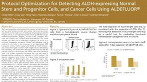 科学海报Protocol Optimization for Detecting ALDH-Expressing Normal Stem and Progenitor Cells and Cancer Cells Using ALDEFLUOR™
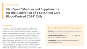 技术公告StemSpan™ Medium and Supplements for the Generation of T Cells from Cord Blood-Derived CD34+ Cells
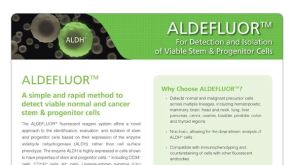 产品手册ALDEFLUOR™: For Identification and Isolation of Viable Stem & Progenitor Cells
 沪公网安备31010102008431号