Lawrence HJ et al. (DEC 2005)
Blood 106 12 3988--94
Loss of expression of the Hoxa-9 homeobox gene impairs the proliferation and repopulating ability of hematopoietic stem cells.
The homeobox gene Hoxa-9 is normally expressed in primitive bone marrow cells,and overexpression of Hoxa-9 markedly expands hematopoietic stem cells,suggesting a function in early hematopoiesis. We present evidence for major functional defects in Hoxa-9-/- hematopoietic stem cells. Hoxa-9-/- marrow cells have normal numbers of immunophenotypic stem cells (Lin(-)c-kit(+)flk-2(-)Sca-1+ [KLFS] cells). However,sublethally irradiated Hoxa-9-/- mice develop persistent pancytopenia,indicating unusual sensitivity to ionizing irradiation. In competitive transplantation assays,Hoxa-9-/- cells showed an 8-fold reduction in multilineage long-term repopulating ability,a defect not seen in marrow cells deficient for the adjacent Hoxa-10 gene. Single-cell cultures of KLFS cells showed a 4-fold reduction in large high-proliferation potential colonies. In liquid cultures,Hoxa-9-deficient Lin(-)Sca-1(+) cells showed slowed proliferation (a 5-fold reduction in cell numbers at day 8) and delayed emergence of committed progenitors (a 5-fold decrease in colony-forming cells). Slowing of proliferation was accompanied by a delay in myeloid maturation,with a decrease in Gr-1hiMac-1hi cells at the end of the culture. Retroviral transduction with a Hoxa-9 expression vector dramatically enhanced the cytokine-driven proliferation and in vivo engraftment of Hoxa-9-/- marrow cells. Hoxa-9 appears to be specifically required for normal hematopoietic stem cell function both in vitro and in vivo.
View Publication
产品类型:
产品号#:
03231
09600
09650
产品名:
MethoCult™M3231
StemSpan™ SFEM
StemSpan™ SFEM
Wu W et al. (JUL 2006)
Blood 108 1 141--51
KSHV/HHV-8 infection of human hematopoietic progenitor (CD34+) cells: persistence of infection during hematopoiesis in vitro and in vivo.
The cellular reservoir for Kaposi sarcoma-associated herpesvirus (KSHV) infection in the hematopoietic compartment and mechanisms governing latent infection and reactivation remain undefined. To determine susceptibility of human CD34+ hematopoietic progenitor cells (HPCs) to infection with KSHV,purified HPCs were exposed to KSHV,and cells were differentiated in vitro and in vivo. Clonogenic colony-forming activity was significantly suppressed in KSHV-infected CD34+ cells,and viral DNA was predominantly localized to granulocyte-macrophage colonies differentiated in vitro. rKSHV.219 is a recombinant KSHV construct that expresses green fluorescent protein from a cellular promoter active during latency and red fluorescent protein from a viral lytic promoter. Infection of CD34+ HPCs with rKSHV.219 showed similar patterns of infection,persistence,and hematopoietic suppression in vitro in comparison with KSHV. rKSHV.219 infection was detected in human CD14+ and CD19+ cells recovered from NOD/SCID mouse bone marrow and spleen following reconstitution with rKSHV.219-infected CD34+ HPCs. These results suggest that rKSHV.219 establishes persistent infection in NOD/SCID mice and that virus may be disseminated following differentiation of infected HPCs into the B-cell and monocyte lineages. CD34+ HPCs may be a reservoir for KSHV infection and may provide a continuous source of virally infected cells in vivo.
View Publication
产品类型:
产品号#:
02690
产品名:
StemSpan™CC100
Milsom MD et al. (MAY 2009)
Blood 113 21 5111--20
Ectopic HOXB4 overcomes the inhibitory effect of tumor necrosis factor-alpha on Fanconi anemia hematopoietic stem and progenitor cells.
Ectopic delivery of HOXB4 elicits the expansion of engrafting hematopoietic stem cells (HSCs). We hypothesized that inhibition of tumor necrosis factor-alpha (TNF-alpha) signaling may be central to the self-renewal signature of HOXB4. Because HSCs derived from Fanconi anemia (FA) knockout mice are hypersensitive to TNF-alpha,we studied Fancc(-/-) HSCs to determine the physiologic effects of HOXB4 on TNF-alpha sensitivity and the relationship of these effects to the engraftment defect of FA HSCs. Overexpression of HOXB4 reversed the in vitro hypersensitivity to TNF-alpha of Fancc(-/-) HSCs and progenitors (P) and partially rescued the engraftment defect of these cells. Coexpression of HOXB4 and the correcting FA-C protein resulted in full correction compared with wild-type (WT) HSCs. Ectopic expression of HOXB4 resulted in a reduction in both apoptosis and reactive oxygen species in Fancc(-/-) but not WT HSC/P. HOXB4 overexpression was also associated with a significant reduction in surface expression of TNF-alpha receptors on Fancc(-/-) HSC/P. Finally,enhanced engraftment was seen even when HOXB4 was expressed in a time-limited fashion during in vivo reconstitution. Thus,the HOXB4 engraftment signature may be related to its effects on TNF-alpha signaling,and this pathway may be a molecular target for timed pharmacologic manipulation of HSC during reconstitution.
View Publication
Multilineage long-term engraftment potential of drug-resistant hematopoietic progenitors.
Peripheral blood progenitor cells (PBPCs) are increasingly used instead of bone marrow for autologous or allogeneic transplantation. In this study PBPCs mobilized in cancer patients by chemotherapy and granulocyte-colony stimulating factor were collected by apheresis and first enriched by immunoaffinity removal of lineage positive cells. When these cells were exposed to both cyclophosphamide and taxol or cultured for 7 days in the presence of 5-fluorouracil,stem cell factor,and interleukin-3,88% to 93% of the enriched PBPCs were killed and short-term clonogenic capacity in methylcellulose assays was lost,but week-5 cobblestone area-forming cell (CAFC) enrichment was higher than 10-fold in comparison to enriched PBPCs and higher than 700-fold in comparison to unmanipulated apheresis cells. After drug exposure,most of the progenitors displayed a CD34+,CD38-,multidrug-resistance (MDR+),Rhodamine 123 low,Hoechst 33342 low phenotype,and as few as 180 of these drug-resistant cells were able to generate a stable multilineage human hematopoiesis in sublethally irradiated immunodeficient mice. In these animals,the level of human hematopoietic engraftment was significantly increased by cotransplantation of irradiated cells from the human L87/4 stromal cell line. These observations are consistent with the functional isolation of a population of very early hematopoietic progenitors and might help to design new protocols for the removal of neoplastic cells from autografts.
View Publication
产品类型:
产品号#:
05150
产品名:
MyeloCult™H5100
Qué et al. (JUN 2011)
Blood 117 22 5918--30
Smad4 binds Hoxa9 in the cytoplasm and protects primitive hematopoietic cells against nuclear activation by Hoxa9 and leukemia transformation.
We studied leukemic stem cells (LSCs) in a Smad4(-/-) mouse model of acute myelogenous leukemia (AML) induced either by the HOXA9 gene or by the fusion oncogene NUP98-HOXA9. Although Hoxa9-Smad4 complexes accumulate in the cytoplasm of normal hematopoietic stem cells and progenitor cells (HSPCs) transduced with these oncogenes,there is no cytoplasmic stabilization of HOXA9 in Smad4(-/-) HSPCs,and as a consequence increased levels of Hoxa9 is observed in the nucleus leading to increased immortalization in vitro. Loss of Smad4 accelerates the development of leukemia in vivo because of an increase in transformation of HSPCs. Therefore,the cytoplasmic binding of Hoxa9 by Smad4 is a mechanism to protect Hoxa9-induced transformation of normal HSPCs. Because Smad4 is a potent tumor suppressor involved in growth control,we developed a strategy to modify the subcellular distribution of Smad4. We successfully disrupted the interaction between Hoxa9 and Smad4 to activate the TGF-β pathway and apoptosis,leading to a loss of LSCs. Together,these findings reveal a major role for Smad4 in the negative regulation of leukemia initiation and maintenance induced by HOXA9/NUP98-HOXA9 and provide strong evidence that antagonizing Smad4 stabilization by these oncoproteins might be a promising novel therapeutic approach in leukemia.
View Publication
产品类型:
产品号#:
03434
03444
03236
产品名:
MethoCult™GF M3434
MethoCult™GF M3434
MethoCult™SF M3236
Conneally E et al. (JAN 1996)
Blood 87 2 456--64
Rapid and efficient selection of human hematopoietic cells expressing murine heat-stable antigen as an indicator of retroviral-mediated gene transfer.
Recombinant retroviruses offer many advantages for the genetic modification of human hematopoietic cells,although their use in clinical protocols has thus far given disappointing results. There is therefore an important need to develop new strategies that will allow effectively transduced primitive hematopoietic target populations to be both rapidly characterized and isolated free of residual nontransduced but biologically equivalent cells. To address this need,we constructed a murine stem cell virus (MSCV)-based retroviral vector containing the 228-bp coding sequence of the murine heat-stable antigen (HSA) and generated helper virus-free amphotropic MSCV-HSA producer cells by transfection of GP-env AM12 packaging cells. Light density and,in some cases,lineage marker-negative (lin-) normal human marrow or mobilized peripheral blood cells preactivated by exposure to interleukin-3 (IL-3),IL-6,and Steel factor in vitro for 48 hours were then infected by cocultivation with these MSCV-HSA producer cells for a further 48 hours in the presence of the same cytokines. Fluorescence-activated cell sorting (FACS) analysis of the cells 24 hours later showed 21% to 41% (mean,27%) of those that were still CD34+ to have acquired the ability to express HSA. The extent of gene transfer to erythroid and granulopoietic progenitors (burst-forming unit-erythroid and colony-forming unit-granulocyte-macrophage),as assessed by the ability of these cells to form colonies of mature progeny in the presence of normally toxic concentrations of G418,averaged 11% and 12%,respectively,in 6 experiments. These values could be increased to 100% and 77%,respectively,by prior isolation of the CD34+HSA+ cell fraction and were correspondingly decreased to an average of 2% and 5%,respectively,in the CD34+HSA- cells. In addition,the extent of gene transfer to long-term culture-initiating cells (LTC-IC) was assessed by G418 resistance. The average gene transfer to LTC-IC-derived colony-forming cells in the unsorted population was textless or = 7% in 4 experiments. FACS selection of the initially CD34+HSA+ cells increased this value to 86% and decreased it to 3% for the LTC-IC plated from the CD34+HSA- cells. Transfer of HSA gene expression to a phenotypically defined more primitive subpopulation of CD34+ cells,ie,those expressing little or no CD38,could also be shown by FACS analysis of infected populations 24 hours after infection. These findings underscore the potential use of retroviral vectors encoding HSA for the specific identification and non-toxic selection immediately after infection of retrovirally transduced populations of primitive human hematopoietic cells. In addition,such vectors should facilitate the subsequent tracking of their marked progeny using multiparameter flow cytometry.
View Publication
产品类型:
产品号#:
04436
04064
04100
04230
04236
04431
04434
04444
04464
04531
04535
04545
04536
04564
04035
04330
04034
04044
04435
04445
04534
04544
04437
04447
产品名:
MethoCult™ SF H4436
MethoCult™ H4034 Optimum启动试剂盒套装
MethoCult™ H4100
MethoCult™H4230
MethoCult™SF H4236
MethoCult™H4431
MethoCult™H4434经典
MethoCult™H4434经典
MethoCult™ H4434 Classic启动试剂盒套装
MethoCult™H4531
MethoCult™H4535富集无EPO
MethoCult™ H4535 Enriched,不含EPO
MethoCult™ SF H4536
入门套件MethoCult™H4534经典无EPO
MethoCult™H4035 Optimum无EPO
MethoCult™H4330
MethoCult™H4034 Optimum
MethoCult™H4034 Optimum
MethoCult™H4435富集
MethoCult™H4435富集
MethoCult™H4534经典无EPO
MethoCult™H4534经典无EPO
MethoCult™表达
MethoCult™表达
(May 2024)
Frontiers in Immunology 15
Single-cell transcriptomic analysis of hematopoietic progenitor cells from patients with systemic lupus erythematosus reveals interferon-inducible reprogramming in early progenitors
IntroductionImmune cells that contribute to the pathogenesis of systemic lupus erythematosus (SLE) derive from adult hematopoietic stem and progenitor cells (HSPCs) within the bone marrow (BM). For this reason,we reasoned that fundamental abnormalities in SLE can be traced to a BM-derived HSPC inflammatory signature.MethodsBM samples from four SLE patients,six healthy controls,and two umbilical cord blood (CB) samples were used. CD34+ cells were isolated from BM and CB samples,and single-cell RNA-sequencing was performed.ResultsA total of 426 cells and 24,473 genes were used in the analysis. Clustering analysis resulted in seven distinct clusters of cell types. Mutually exclusive markers,which were characteristic of each cell type,were identified. We identified three HSPC subpopulations,one of which consisted of proliferating cells (MKI67 expressing cells),one T-like,one B-like,and two myeloid-like progenitor subpopulations. Differential expression analysis revealed i) cell cycle-associated signatures,in healthy BM of HSPC clusters 3 and 4 when compared with CB,and ii) interferon (IFN) signatures in SLE BM of HSPC clusters 3 and 4 and myeloid-like progenitor cluster 5 when compared with healthy controls. The IFN signature in SLE appeared to be deregulated following TF regulatory network analysis and differential alternative splicing analysis between SLE and healthy controls in HSPC subpopulations.DiscussionThis study revealed both quantitative—as evidenced by decreased numbers of non-proliferating early progenitors—and qualitative differences—characterized by an IFN signature in SLE,which is known to drive loss of function and depletion of HSPCs. Chronic IFN exposure affects early hematopoietic progenitors in SLE,which may account for the immune aberrancies and the cytopenias in SLE.
View Publication
产品类型:
产品号#:
17856
17856RF
100-1569
产品名:
EasySep™人CD34正选试剂盒 II
EasySep™人CD34正选试剂盒 II
EasySep™人CD34正选试剂盒 II
P. Singh et al. (may 2020)
Stem cell reviews and reports
Aging-Related Reduced Expression of CXCR4 on Bone Marrow Mesenchymal Stromal Cells Contributes to Hematopoietic Stem and Progenitor Cell Defects.
Aging impairs the regenerative potential of hematopoietic stem cells (HSC) and skews differentiation towards the myeloid lineage. The bone marrow (BM) microenvironment has recently been suggested to influence HSC aging,however the mechanisms whereby BM stromal cells mediate this effect is unknown. Here we show that aging-associated decreased expression of CXCR4 expression on BM mesenchymal stem cells (MSC) plays a crucial role in the development of the hematopoietic stem and progenitor cells (HSPC) aging phenotype. The BM MSC from old mice was sufficient to drive a premature aging phenotype of young HSPC when cultured together ex vivo. The impaired ability of old MSC to support HSPC function is associated with reduced expression of CXCR4 on BM MSC of old mice. Deletion of the CXCR4 gene in young MSC accelerates an aging phenotype in these cells characterized by increased production of reactive oxygen species (ROS),DNA damage,senescence,and reduced proliferation. Culture of HSPC from young mice with CXCR4 deficient MSC also from young mice led to a premature aging phenotype in the young HSPC,as evidenced by reduced hematopoietic regeneration and enhanced myeloid differentiation. Mechanistically,CXCR4 signaling prevents BM MSC dysfunction by suppressing oxidative stress,as treatment of old or CXCR4 deficient MSC with N-acetyl-L-cysteine (NAC),improved their niche supporting activity,and attenuated the HSPC aging phenotype. Our studies suggest that age-associated reduction in CXCR4 expression on BM MSC impairs hematopoietic niche activity with increased ROS production,driving an HSC aging phenotype. Thus,modulation of the SDF-1/CXCR4 axis in MSC may lead to novel interventions to alleviate the age-associated decline in immune/hematopoietic function.
View Publication
产品类型:
产品号#:
05513
产品名:
MesenCult™ 扩增试剂盒 (小鼠)
Kamminga LM et al. (MAR 2006)
Blood 107 5 2170--9
The Polycomb group gene Ezh2 prevents hematopoietic stem cell exhaustion.
The molecular mechanism responsible for a decline of stem cell functioning after replicative stress remains unknown. We used mouse embryonic fibroblasts (MEFs) and hematopoietic stem cells (HSCs) to identify genes involved in the process of cellular aging. In proliferating and senescent MEFs one of the most differentially expressed transcripts was Enhancer of zeste homolog 2 (Ezh2),a Polycomb group protein (PcG) involved in histone methylation and deacetylation. Retroviral overexpression of Ezh2 in MEFs resulted in bypassing of the senescence program. More importantly,whereas normal HSCs were rapidly exhausted after serial transplantations,overexpression of Ezh2 completely conserved long-term repopulating potential. Animals that were reconstituted with 3 times serially transplanted control bone marrow cells all died due to hematopoietic failure. In contrast,similarly transplanted Ezh2-overexpressing stem cells restored stem cell quality to normal levels. In a genetic genomics" screen�
View Publication

 EasySep™小鼠TIL(CD45)正选试剂盒


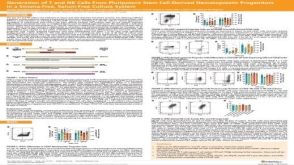 科学海报Generation of T and NK Cells From Pluripotent Stem Cell-Derived Hematopoietic Progenitors in a Stroma-Free, Serum-Free Culture System
 挂图Identification of Colonies Derived from Human Hematopoietic Progenitors Representative colony images and tips for identifying progenitor subtypes in CFU assays
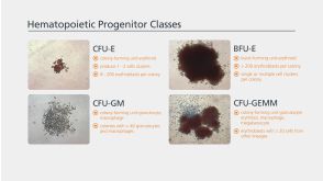

 沪公网安备31010102008431号