Nottingham WT et al. (DEC 2007)
Blood 110 13 4188--97
Runx1-mediated hematopoietic stem-cell emergence is controlled by a Gata/Ets/SCL-regulated enhancer.
The transcription factor Runx1/AML1 is an important regulator of hematopoiesis and is critically required for the generation of the first definitive hematopoietic stem cells (HSCs) in the major vasculature of the mouse embryo. As a pivotal factor in HSC ontogeny,its transcriptional regulation is of high interest but is largely undefined. In this study,we used a combination of comparative genomics and chromatin analysis to identify a highly conserved 531-bp enhancer located at position + 23.5 in the first intron of the 224-kb mouse Runx1 gene. We show that this enhancer contributes to the early hematopoietic expression of Runx1. Transcription factor binding in vivo and analysis of the mutated enhancer in transient transgenic mouse embryos implicate Gata2 and Ets proteins as critical factors for its function. We also show that the SCL/Lmo2/Ldb-1 complex is recruited to the enhancer in vivo. Importantly,transplantation experiments demonstrate that the intronic Runx1 enhancer targets all definitive HSCs in the mouse embryo,suggesting that it functions as a crucial cis-regulatory element that integrates the Gata,Ets,and SCL transcriptional networks to initiate HSC generation.
View Publication
BACKGROUND & AIMS The early embryonic pancreas gives rise to exocrine (ducts and acini) and endocrine lineages. Control of exocrine differentiation is poorly understood,but may be a critical avenue through which to manipulate pancreatic ductal carcinoma. Retinoids have been shown to change the character of pancreatic ductal cancer cells to a less malignant phenotype. We have shown that 9-cis retinoic acid (9cRA) inhibits acinar differentiation in the developing pancreas,in favor of ducts,and we wanted to determine the role of retinoids in duct versus acinar differentiation. METHODS We used multiple culture systems for the 11-day embryonic mouse pancreas. RESULTS Retinoic acid receptor (RAR)-selective agonists mimicked the acinar suppressive effect of 9cRA,suggesting that RAR-RXR heterodimers were critical to ductal differentiation. RARalpha was only expressed in mesenchyme,whereas RXRalpha was expressed in epithelium and mesenchyme. Retinaldehyde dehydrogenase 2,a critical enzyme in retinoid synthesis,was expressed only in pancreatic epithelium. 9cRA did not induce ductal differentiation in the absence of mesenchyme,implicating a requirement for mesenchyme in 9cRA effects. Mesenchymal laminin is necessary for duct differentiation,and retinoids are known to enhance laminin expression. In 9cRA-treated pancreas,immunohistochemistry for laminin showed a strong band of staining around ducts,and blockage of laminin signaling blocked all 9cRA effects. Western blot and RT-PCR of pancreatic mesenchyme showed laminin-beta1 protein and mRNA induction by 9cRA. CONCLUSIONS Retinoids regulate exocrine lineage selection through epithelial-mesenchymal interactions,mediated through up-regulation of mesenchymal laminin-1.
View Publication
产品类型:
产品号#:
72382
产品名:
9-cis Retinoic Acid
Aranha M et al. (JAN 2010)
BMC genomics 11 514
Apoptosis-associated microRNAs are modulated in mouse, rat and human neural differentiation.
BACKGROUND MicroRNAs (miRs or miRNAs) regulate several biological processes in the cell. However,evidence for miRNAs that control the differentiation program of specific neural cell types has been elusive. Recently,we have shown that apoptosis-associated factors,such as p53 and caspases participate in the differentiation process of mouse neural stem (NS) cells. To identify apoptosis-associated miRNAs that might play a role in neuronal development,we performed global miRNA expression profiling experiments in NS cells. Next,we characterized the expression of proapoptotic miRNAs,including miR-16,let-7a and miR-34a in distinct models of neural differentiation,including mouse embryonic stem cells,PC12 and NT2N cells. In addition,the expression of antiapoptotic miR-19a and 20a was also evaluated. RESULTS The expression of miR-16,let-7a and miR-34a was consistently upregulated in neural differentiation models. In contrast,expression of miR-19a and miR-20a was downregulated in mouse NS cell differentiation. Importantly,differential expression of specific apoptosis-related miRNAs was not associated with increased cell death. Overexpression of miR-34a increased the proportion of postmitotic neurons of mouse NS cells. CONCLUSIONS In conclusion,the identification of miR-16,let-7a and miR-34a,whose expression patterns are conserved in mouse,rat and human neural differentiation,implicates these specific miRNAs in mammalian neuronal development. The results provide new insights into the regulation of neuronal differentiation by apoptosis-associated miRNAs.
View Publication
Hypoxic stress underlies defects in erythroblast islands in the Rb-null mouse.
Definitive erythropoiesis occurs in islands composed of a central macrophage in contact with differentiating erythroblasts. Erythroid maturation including enucleation can also occur in the absence of macrophages both in vivo and in vitro. We reported previously that loss of Rb induces cell-autonomous defects in red cell maturation under stress conditions,while other reports have suggested that the failure of Rb-null erythroblasts to enucleate is due to defects in associated macrophages. Here we show that erythropoietic islands are disrupted by hypoxic stress,such as occurs in the Rb-null fetal liver,that Rb(-/-) macrophages are competent for erythropoietic island formation in the absence of exogenous stress and that enucleation defects persist in Rb-null erythroblasts irrespective of macrophage function.
View Publication
产品类型:
产品号#:
03434
03444
09600
09650
产品名:
MethoCult™GF M3434
MethoCult™GF M3434
StemSpan™ SFEM
StemSpan™ SFEM
Garaycoechea JI et al. (SEP 2012)
Nature 489 7417 571--5
Genotoxic consequences of endogenous aldehydes on mouse haematopoietic stem cell function.
Haematopoietic stem cells (HSCs) regenerate blood cells throughout the lifespan of an organism. With age,the functional quality of HSCs declines,partly owing to the accumulation of damaged DNA. However,the factors that damage DNA and the protective mechanisms that operate in these cells are poorly understood. We have recently shown that the Fanconi anaemia DNA-repair pathway counteracts the genotoxic effects of reactive aldehydes. Mice with combined inactivation of aldehyde catabolism (through Aldh2 knockout) and the Fanconi anaemia DNA-repair pathway (Fancd2 knockout) display developmental defects,a predisposition to leukaemia,and are susceptible to the toxic effects of ethanol-an exogenous source of acetaldehyde. Here we report that aged Aldh2(-/-) Fancd2(-/-) mutant mice that do not develop leukaemia spontaneously develop aplastic anaemia,with the concomitant accumulation of damaged DNA within the haematopoietic stem and progenitor cell (HSPC) pool. Unexpectedly,we find that only HSPCs,and not more mature blood precursors,require Aldh2 for protection against acetaldehyde toxicity. Additionally,the aldehyde-oxidizing activity of HSPCs,as measured by Aldefluor stain,is due to Aldh2 and correlates with this protection. Finally,there is more than a 600-fold reduction in the HSC pool of mice deficient in both Fanconi anaemia pathway-mediated DNA repair and acetaldehyde detoxification. Therefore,the emergence of bone marrow failure in Fanconi anaemia is probably due to aldehyde-mediated genotoxicity restricted to the HSPC pool. These findings identify a new link between endogenous reactive metabolites and DNA damage in HSCs,and define the protective mechanisms that counteract this threat.
View Publication
Hisa T et al. (JAN 2004)
The EMBO journal 23 2 450--9
Hematopoietic, angiogenic and eye defects in Meis1 mutant animals.
Meis1 and Hoxa9 expression is upregulated by retroviral integration in murine myeloid leukemias and in human leukemias carrying MLL translocations. Both genes also cooperate to induce leukemia in a mouse leukemia acceleration assay,which can be explained,in part,by their physical interaction with each other as well as the PBX family of homeodomain proteins. Here we show that Meis1-deficient embryos have partially duplicated retinas and smaller lenses than normal. They also fail to produce megakaryocytes,display extensive hemorrhaging,and die by embryonic day 14.5. In addition,Meis1-deficient embryos lack well-formed capillaries,although larger blood vessels are normal. Definitive myeloerythroid lineages are present in the mutant embryos,but the total numbers of colony-forming cells are dramatically reduced. Mutant fetal liver cells also fail to radioprotect lethally irradiated animals and they compete poorly in repopulation assays even though they can repopulate all hematopoietic lineages. These and other studies showing that Meis1 is expressed at high levels in hematopoietic stem cells (HSCs) suggest that Meis1 may also be required for the proliferation/self-renewal of the HSC.
View Publication
产品类型:
产品号#:
04960
04902
04900
04961
04901
04963
04962
04970
04971
产品名:
MegaCult™-C胶原蛋白和不含细胞因子的培养基
胶原蛋白溶液
MegaCult™-C培养基无细胞因子
MegaCult™-C胶原蛋白和细胞因子培养基
MegaCult™-C细胞因子培养基
双室载玻片试剂盒
MegaCult™-C cfu染色试剂盒
MegaCult™-C不含细胞因子完整试剂盒
MegaCult™-C细胞因子完整试剂盒
Ragu C et al. (NOV 2010)
Blood 116 22 4464--73
The transcription factor Srf regulates hematopoietic stem cell adhesion.
Adhesion properties of hematopoietic stem cells (HSCs) in the bone marrow (BM) niches control their migration and affect their cell-cycle dynamics. The serum response factor (Srf) regulates growth factor-inducible genes and genes controlling cytoskeleton structures involved in cell spreading,adhesion,and migration. We identified a role for Srf in HSC adhesion and steady-state hematopoiesis. Conditional deletion of Srf in BM cells resulted in a 3-fold expansion of the long- and short-term HSCs and multipotent progenitors (MPPs),which occurs without long-term modification of cell-cycle dynamics. Early differentiation steps to myeloid and lymphoid lineages were normal,but Srf loss results in alterations in mature-cell production and severe thrombocytopenia. Srf-null BM cells also displayed compromised engraftment properties in transplantation assays. Gene expression analysis identified Srf target genes expressed in HSCs,including a network of genes associated with cell migration and adhesion. Srf-null stem cells and MPPs displayed impair expression of the integrin network and decreased adherence in vitro. In addition,Srf-null mice showed increase numbers of circulating stem and progenitor cells,which likely reflect their reduced retention in the BM. Altogether,our results demonstrate that Srf is an essential regulator of stem cells and MPP adhesion,and suggest that Srf acts mainly through cell-matrix interactions and integrin signaling.
View Publication

 EasySep™小鼠TIL(CD45)正选试剂盒


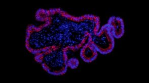 实验方案How to Establish Intestinal Organoid Culture from Isolated Mouse Intestinal Crypts
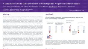 科学海报Fast and Easy Hematopoietic Progenitor Cell Enrichment With SepMate™


 沪公网安备31010102008431号