M. E. Stremska et al. (may 2019)
Journal of autoimmunity
IL233, an IL-2-IL-33 hybrid cytokine induces prolonged remission of mouse lupus nephritis by targeting Treg cells as a single therapeutic agent.
Lupus glomerulonephritis (GN) is an autoimmune disease characterized by immune complex-deposition,complement activation and glomerular inflammation. In lupus-prone NZM2328 mice,the occurrence of lupus GN was accompanied by a decrease in Treg cells and an increase in proinflammatory cytokine-producing T cells. Because IL-33 in addition to IL-2 has been shown to be important for Treg cell proliferation and ST2 (IL-33 receptor) positive Treg cells are more potent in suppressor activity,a hybrid cytokine with active domains of IL-2 and IL-33 was generated to target the ST2+ Treg cells as a therapeutic agent to treat lupus GN. Three mouse models were used: spontaneous and Ad-IFNalpha- accelerated lupus GN in NZM2328 and the lymphoproliferative autoimmune GN in MRL/lpr mice. Daily injections of IL233 for 5 days prevented Ad-IFNalpha-induced lupus GN and induced remission of spontaneous lupus GN. The remission was permanent in that no relapses were detected. The remission was accompanied by persistent elevation of Treg cells in the renal lymph nodes. IL233 is more potent than IL-2 and IL-33 either singly or in combination in the treatment of lupus GN. The results of this study support the thesis that IL233 should be considered as a novel agent for treating lupus GN.
View Publication
D. Kobayashi et al. ( 2022)
Frontiers in immunology 13 973880
Tas2R signaling enhances mouse neutrophil migration via a ROCK-dependent pathway.
Type-2 bitter taste receptors (Tas2Rs) are a large family of G protein-coupled receptors that are expressed in the oral cavity and serve to detect substances with bitter tastes in foods and medicines. Recent evidence suggests that Tas2Rs are also expressed extraorally,including in immune cells. However,the role of Tas2Rs in immune cells remains controversial. Here,we demonstrate that Tas2R126,Tas2R135,and Tas2R143 are expressed in mouse neutrophils,but not in other immune cells such as macrophages or T and B lymphocytes. Treatment of bone marrow-derived neutrophils from wild-type mice with the Tas2R126/143 agonists arbutin and d-salicin led to enhanced C-X-C motif chemokine ligand 2 (CXCL2)-stimulated migration in vitro,but this response was not observed in neutrophils from Tas2r126/135/143-deficient mice. Enhancement of CXCL2-stimulated migration by Tas2R agonists was accompanied by increased phosphorylation of myosin light chain 2 (MLC2) and was blocked by pretreatment of neutrophils with inhibitors of Rho-associated coiled-coil-containing protein kinase (ROCK),but not by inhibitors of the small GTPase RhoA. Taken together,these results demonstrate that mouse neutrophils express functional Tas2R126/143 and suggest a role for Tas2R126/143-ROCK-MLC2-dependent signaling in the regulation of neutrophil migration.
View Publication
Reconstitution of the functional human hematopoietic microenvironment derived from human mesenchymal stem cells in the murine bone marrow compartment.
Hematopoiesis is maintained by specific interactions between both hematopoietic and nonhematopoietic cells. Whereas hematopoietic stem cells (HSCs) have been extensively studied both in vitro and in vivo,little is known about the in vivo characteristics of stem cells of the nonhematopoietic component,known as mesenchymal stem cells (MSCs). Here we have visualized and characterized human MSCs in vivo following intramedullary transplantation of enhanced green fluorescent protein-marked human MSCs (eGFP-MSCs) into the bone marrow (BM) of nonobese diabetic/severe combined immunodeficiency (NOD/SCID) mice. Between 4 to 10 weeks after transplantation,eGFP-MSCs that engrafted in murine BM integrated into the hematopoietic microenvironment (HME) of the host mouse. They differentiated into pericytes,myofibroblasts,BM stromal cells,osteocytes in bone,bone-lining osteoblasts,and endothelial cells,which constituted the functional components of the BM HME. The presence of human MSCs in murine BM resulted in an increase in functionally and phenotypically primitive human hematopoietic cells. Human MSC-derived cells that reconstituted the HME appeared to contribute to the maintenance of human hematopoiesis by actively interacting with primitive human hematopoietic cells.
View Publication
产品类型:
产品号#:
04034
04044
产品名:
MethoCult™H4034 Optimum
MethoCult™H4034 Optimum
Naramura M et al. (SEP 2010)
Proceedings of the National Academy of Sciences of the United States of America 107 37 16274--9
Rapidly fatal myeloproliferative disorders in mice with deletion of Casitas B-cell lymphoma (Cbl) and Cbl-b in hematopoietic stem cells.
Casitas B-cell lymphoma (Cbl)-family E3 ubiquitin ligases are negative regulators of tyrosine kinase signaling. Recent work has revealed a critical role of Cbl in the maintenance of hematopoietic stem cell (HSC) homeostasis,and mutations in CBL have been identified in myeloid malignancies. Here we show that,in contrast to Cbl or Cbl-b single-deficient mice,concurrent loss of Cbl and Cbl-b in the HSC compartment leads to an early-onset lethal myeloproliferative disease in mice. Cbl,Cbl-b double-deficient bone marrow cells are hypersensitive to cytokines,and show altered biochemical response to thrombopoietin. Thus,Cbl and Cbl-b play redundant but essential roles in HSC regulation,whose breakdown leads to hematological abnormalities that phenocopy crucial aspects of mutant Cbl-driven human myeloid malignancies.
View Publication
产品类型:
产品号#:
03234
产品名:
MethoCult™M3234
Christopher MJ et al. (FEB 2011)
The Journal of experimental medicine 208 2 251--60
Expression of the G-CSF receptor in monocytic cells is sufficient to mediate hematopoietic progenitor mobilization by G-CSF in mice.
Granulocyte colony-stimulating factor (G-CSF),the prototypical mobilizing cytokine,induces hematopoietic stem and progenitor cell (HSPC) mobilization from the bone marrow in a cell-nonautonomous fashion. This process is mediated,in part,through suppression of osteoblasts and disruption of CXCR4/CXCL12 signaling. The cellular targets of G-CSF that initiate the mobilization cascade have not been identified. We use mixed G-CSF receptor (G-CSFR)-deficient bone marrow chimeras to show that G-CSF-induced mobilization of HSPCs correlates poorly with the number of wild-type neutrophils. We generated transgenic mice in which expression of the G-CSFR is restricted to cells of the monocytic lineage. G-CSF-induced HSPC mobilization,osteoblast suppression,and inhibition of CXCL12 expression in the bone marrow of these transgenic mice are intact,demonstrating that G-CSFR signals in monocytic cells are sufficient to induce HSPC mobilization. Moreover,G-CSF treatment of wild-type mice is associated with marked loss of monocytic cells in the bone marrow. Finally,we show that bone marrow macrophages produce factors that support the growth and/or survival of osteoblasts in vitro. Together,these data suggest a model in which G-CSFR signals in bone marrow monocytic cells inhibit the production of trophic factors required for osteoblast lineage cell maintenance,ultimately leading to HSPC mobilization.
View Publication
产品类型:
产品号#:
03434
03444
产品名:
MethoCult™GF M3434
MethoCult™GF M3434
S. H. Park et al. (may 2019)
Nucleic acids research
Highly efficient editing of the beta-globin gene in patient-derived hematopoietic stem and progenitor cells to treat sickle cell disease.
Sickle cell disease (SCD) is a monogenic disorder that affects millions worldwide. Allogeneic hematopoietic stem cell transplantation is the only available cure. Here,we demonstrate the use of CRISPR/Cas9 and a short single-stranded oligonucleotide template to correct the sickle mutation in the beta-globin gene in hematopoietic stem and progenitor cells (HSPCs) from peripheral blood or bone marrow of patients with SCD,with 24.5 ± 7.6{\%} efficiency without selection. Erythrocytes derived from gene-edited cells showed a marked reduction of sickle cells,with the level of normal hemoglobin (HbA) increased to 25.3 ± 13.9{\%}. Gene-corrected SCD HSPCs retained the ability to engraft when transplanted into non-obese diabetic (NOD)-SCID-gamma (NSG) mice with detectable levels of gene correction 16-19 weeks post-transplantation. We show that,by using a high-fidelity SpyCas9 that maintained the same level of on-target gene modification,the off-target effects including chromosomal rearrangements were significantly reduced. Taken together,our results demonstrate efficient gene correction of the sickle mutation in both peripheral blood and bone marrow-derived SCD HSPCs,a significant reduction in sickling of red blood cells,engraftment of gene-edited SCD HSPCs in vivo and the importance of reducing off-target effects; all are essential for moving genome editing based SCD treatment into clinical practice.
View Publication
产品类型:
产品号#:
04434
04444
产品名:
MethoCult™H4434经典
MethoCult™H4434经典
(Mar 2025)
Frontiers in Immunology 16 19
Characterization of TLR9 responsiveness in cell subsets derived from in vitro pDC differentiation of hematopoietic stem and progenitor cells
Plasmacytoid dendritic cells (pDCs) are multifunctional immune cells with roles in both the innate and adaptive immune system. Their hallmark function is production of large amounts of type I interferons in response to viral infections,but they are also capable of producing a range of other cytokines,antigen presentation,and cytotoxicity. Their potential as an immunotherapy for cancer and infectious disease is being explored,but broad application of these cells is challenged by low frequency in the blood and low viability during ex vivo culturing. We have previously developed an effective in vitro differentiation protocol for producing pDCs from CD34+ hematopoietic stem and progenitor cells (HSPC-pDCs),which provides an attainable and large source of pDCs. HSPC-pDCs present pDC characteristics and functions,and like naturally occurring pDCs they exhibit large phenotypic and functional heterogeneity. Here,we characterize different cell subsets from in vitro pDC differentiation and identify a distinct population,which is the major producer of IFNα in response to TLR9 stimulation and display a transcriptomic profile similar to what is seen for pDCs circulating in the blood. We also investigate the possibility of rerouting subset specification during HSPCs-to-pDC differentiation by controlling gene expression of key master transcription factors (TFs). We identify TFs associated with the pDC differentiation trajectory that are essential for the development of TLR9-responsive HSPC-pDCs,and we also identify TFs that increase their frequency. In conclusion,we phenotypically and functionally characterize different cell subsets and modulate their relative frequencies by manipulating TF expression during pDC differentiation. These findings provide a deeper understanding of in vitro-differentiated pDC cultures that may spur further developments in their use as an immunomodulatory cell therapy.
View Publication
Titmarsh D et al. (DEC 2011)
Biotechnology and Bioengineering 108 12 2894--2904
Optimization of flowrate for expansion of human embryonic stem cells in perfusion microbioreactors.
Microfluidic systems create significant opportunities to establish highly controlled microenvironmental conditions for screening pluripotent stem cell fate. However,since cell fate is crucially dependent on this microenvironment,it remains unclear as to whether continual perfusion of culture medium supports pluripotent stem cell maintenance in feeder-free,chemically defined conditions,and further,whether optimum perfusion conditions exist for subsequent use of human embryonic stem cell (hESCs) in other microfludic systems. To investigate this,we designed microbioreactors based on resistive flow to screen hESCs under a linear range of flowrates. We report that at low rates (conditions where glucose transport is convection-limited with Péclet number textless1),cells are affected by apparent nutrient depletion and waste accumulation,evidenced by reduced cell expansion and altered morphology. At higher rates,cells are spontaneously washed out,and display morphological changes which may be indicative of early-stage differentiation. However,between these thresholds exists a narrow range of flowrates in which hESCs expand comparably to the equivalent static culture system,with regular morphology and maintenance of the pluripotency marker TG30 in textgreater95% of cells over 7 days. For MEL1 hESCs the optimum flowrate also coincided with the time-averaged medium exchange rate in static cultures,which may therefore provide a good first estimate of appropriate perfusion rates. Overall,we demonstrate hESCs can be maintained in microbioreactors under continual flow for up to 7 days,a critical outcome for the future development of microbioreactor-based screening systems and assays for hESC culture.
View Publication

 EasySep™小鼠TIL(CD45)正选试剂盒


 科学海报Immunomagnetic Cell Enrichment of Lymphoid Progenitors from Mouse Bone Marrow
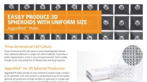 产品手册AggreWell™ for Spheroids
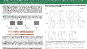 科学海报Defined Culture Conditions for Efficient Derivation Expansion and Cryopreservation of Mesenchymal Progenitor Cells from Human Pluripotent Stem Cells

 沪公网安备31010102008431号