
 EasySep™小鼠TIL(CD45)正选试剂盒


搜索结果: 'methocult media formulations for human hematopoietic cells serum containing'
-

产品类型:
产品号#:
18000
产品名:
EasySep™磁极
-
产品类型:
产品号#:
05850
05857
05870
05875
85850
85857
85870
85875
产品名:
mTeSR™1
mTeSR™1
-
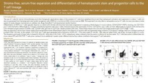 科学海报Stroma-Free, Serum-Free Expansion and Differentiation of Hematopoietic Stem and Progenitor Cells to the T Cell Lineage
产品类型:
Conference:
AAi 2017
产品号#:
09605
09900
09910
09920
产品名:
StemSpan™ SFEM II
-
产品类型:
产品号#:
07801
07811
07851
07861
85450
85460
86450
86460
18060
18061
产品名:
Lymphoprep™
Lymphoprep™
SepMate™-50 (IVD)
SepMate™-50 (IVD)
SepMate™-50 (RUO)
SepMate™-50 (RUO)
Lymphoprep™
Lymphoprep™
-
产品类型:
产品号#:
100-0352
产品名:
条件性重编程(CR)培养基
-
产品类型:
产品号#:
05850
05857
05870
05875
85850
85857
85870
85875
产品名:
mTeSR™1
mTeSR™1
-
产品类型:
产品号#:
05850
05857
05870
05875
85850
85857
85870
85875
产品名:
mTeSR™1
mTeSR™1
-
产品类型:
产品号#:
03434
03444
产品名:
MethoCult™ GF M3434
MethoCult™ GF M3434
-
产品类型:
产品号#:
05150
05350
产品名:
MyeloCult™ H5100
-
产品类型:
产品号#:
09600
09650
28600
产品名:
StemSpan™ SFEM
StemSpan™ SFEM
L-Calc™有限稀释软件
-
产品类型:
产品号#:
产品名:


 沪公网安备31010102008431号