Haploinsufficiency for ribosomal protein genes causes selective activation of p53 in human erythroid progenitor cells.
Haploinsufficiency for ribosomal protein genes has been implicated in the pathophysiology of Diamond-Blackfan anemia (DBA) and the 5q-syndrome,a subtype of myelodysplastic syndrome. The p53 pathway is activated by ribosome dysfunction,but the molecular basis for selective impairment of the erythroid lineage in disorders of ribosome function has not been determined. We found that p53 accumulates selectively in the erythroid lineage in primary human hematopoietic progenitor cells after expression of shRNAs targeting RPS14,the ribosomal protein gene deleted in the 5q-syndrome,or RPS19,the most commonly mutated gene in DBA. Induction of p53 led to lineage-specific accumulation of p21 and consequent cell cycle arrest in erythroid progenitor cells. Pharmacologic inhibition of p53 rescued the erythroid defect,whereas nutlin-3,a compound that activates p53 through inhibition of HDM2,selectively impaired erythropoiesis. In bone marrow biopsies from patients with DBA or del(5q) myelodysplastic syndrome,we found an accumulation of nuclear p53 staining in erythroid progenitor cells that was not present in control samples. Our findings indicate that the erythroid lineage has a low threshold for the induction of p53,providing a basis for the failure of erythropoiesis in the 5q-syndrome,DBA,and perhaps other bone marrow failure syndromes.
View Publication
产品类型:
产品号#:
03334
03434
03444
产品名:
MethoCult™M3334
MethoCult™GF M3434
MethoCult™GF M3434
Liang Y et al. (APR 2013)
Chinese journal of cancer 32 4 205--12
The propensity for tumorigenesis in human induced pluripotent stem cells is related with genomic instability.
The discovery of induced pluripotent stem cells(iPSCs) is a promising advancement in the field of regenerative medicine. Previous studies have indicated that the teratoma-forming propensity of iPSCs is variable; however,the relationship between tumorigenic potential and genomic instability in human iPSCs (HiPSCs) remains to be fully elucidated. Here,we evaluated the malignant potential of HiPSCs by using both colony formation assays and tumorigenicity tests. We demonstrated that HiPSCs formed tumorigenic colonies when grown in cancer cell culture medium and produced malignancies in immunodeficient mice. Furthermore,we analyzed genomic instability in HiPSCs using whole-genome copy number variation analysis and determined that the extent of genomic instability was related with both the cells' propensity to form colonies and their potential for tumorigenesis. These findings indicate a risk for potential malignancy of HiPSCs derived from genomic instability and suggest that quality control tests,including comprehensive tumorigenicity assays and genomic integrity validation,should be rigorously executed before the clinical application of HiPSCs. In addition,HiPSCs should be generated through the use of combined factors or other approaches that decrease the likelihood of genomic instability.
View Publication
产品类型:
产品号#:
85850
85857
产品名:
mTeSR™1
mTeSR™1
Burgos-Ojeda D et al. (JUN 2013)
Cancer research 73 12 3555--3565
A novel model for evaluating therapies targeting human tumor vasculature and human cancer stem-like cells.
Human tumor vessels express tumor vascular markers (TVM),proteins that are not expressed in normal blood vessels. Antibodies targeting TVMs could act as potent therapeutics. Unfortunately,preclinical in vivo studies testing anti-human TVM therapies have been difficult to do due to a lack of in vivo models with confirmed expression of human TVMs. We therefore evaluated TVM expression in a human embryonic stem cell-derived teratoma (hESCT) tumor model previously shown to have human vessels. We now report that in the presence of tumor cells,hESCT tumor vessels express human TVMs. The addition of mouse embryonic fibroblasts and human tumor endothelial cells significantly increases the number of human tumor vessels. TVM induction is mostly tumor-type-specific with ovarian cancer cells inducing primarily ovarian TVMs,whereas breast cancer cells induce breast cancer specific TVMs. We show the use of this model to test an anti-human specific TVM immunotherapeutics; anti-human Thy1 TVM immunotherapy results in central tumor necrosis and a three-fold reduction in human tumor vascular density. Finally,we tested the ability of the hESCT model,with human tumor vascular niche,to enhance the engraftment rate of primary human ovarian cancer stem-like cells (CSC). ALDH(+) CSC from patients (n = 6) engrafted in hESCT within 4 to 12 weeks whereas none engrafted in the flank. ALDH(-) ovarian cancer cells showed no engraftment in the hESCT or flank (n = 3). Thus,this model represents a useful tool to test anti-human TVM therapy and evaluate in vivo human CSC tumor biology.
View Publication
产品类型:
产品号#:
01700
01705
01702
产品名:
ALDEFLUOR™ 试剂盒
ALDEFLUOR™ DEAB试剂
ALDEFLUOR™测定缓冲液
(Sep 2024)
Scientific Reports 14
Generation of a pancreas derived hydrogel for the culture of hiPSC derived pancreatic endocrine cells
Stem cell-derived ?-cells (SC-BCs) represent a potential source for curing diabetes. To date,in vitro generated SC-BCs display an immature phenotype and lack important features in comparison to their bona-fide counterparts. Transplantation into a living animal promotes SC-BCs maturation,indicating that components of the in vivo microenvironment trigger final SC-BCs development. Here,we investigated whether cues of the pancreas specific extracellular matrix (ECM) can improve the differentiation of human induced pluripotent stem cells (hiPSCs) towards ?-cells in vitro. To this aim,a pancreas specific ECM (PanMa) hydrogel was generated from decellularized porcine pancreas and its effect on the differentiation of hiPSC-derived pancreatic hormone expressing cells (HECs) was tested. The hydrogel solidified upon neutralization at 37 °C with gelation kinetics similar to Matrigel. Cytocompatibility of the PanMa hydrogel was demonstrated for a culture duration of 21 days. Encapsulation and culture of HECs in the PanMa hydrogel over 7 days resulted in a stable gene and protein expression of most ?-cell markers,but did not improve ?-cell identity. In conclusion,the study describes the production of a PanMa hydrogel,which provides the basis for the development of ECM hydrogels that are more adapted to the demands of SC-BCs.
View Publication
de Boer AS et al. (AUG 2014)
Science Translational Medicine 6 248 248ra104--248ra104
Genetic validation of a therapeutic target in a mouse model of ALS
AbstractBack to TopbackslashnNeurons produced from stem cells have emerged as a tool to identify new therapeutic targets for neurological diseases such as amyotrophic lateral sclerosis (ALS). However,it remains unclear to what extent these new mechanistic insights will translate to animal models,an important step in the validation of new targets. Previously,we found that glia from mice carrying the SOD1G93A mutation,a model of ALS,were toxic to stem cell–derived human motor neurons. We use pharmacological and genetic approaches to demonstrate that the prostanoid receptor DP1 mediates this glial toxicity. Furthermore,we validate the importance of this mechanism for neural degeneration in vivo. Genetic ablation of DP1 in SOD1G93A mice extended life span,decreased microglial activation,and reduced motor neuron loss. Our findings suggest that blocking DP1 may be a therapeutic strategy in ALS and demonstrate that discoveries from stem cell models of disease can be corroborated in vivo.
View Publication
产品类型:
产品号#:
85850
85857
产品名:
mTeSR™1
mTeSR™1
J. Min et al. (dec 2019)
Nature communications 10 1 5549
Heterogeneity and dynamics of active Kras-induced dysplastic lineages from mouse corpus stomach.
Dysplasia is considered a key transition state between pre-cancer and cancer in gastric carcinogenesis. However,the cellular or phenotypic heterogeneity and mechanisms of dysplasia progression have not been elucidated. We have established metaplastic and dysplastic organoid lines,derived from Mist1-Kras(G12D) mouse stomach corpus and studied distinct cellular behaviors and characteristics of metaplastic and dysplastic organoids. We also examined functional roles for Kras activation in dysplasia progression using Selumetinib,a MEK inhibitor,which is a downstream mediator of Kras signaling. Here,we report that dysplastic organoids die or show altered cellular behaviors and diminished aggressive behavior in response to MEK inhibition. However,the organoids surviving after MEK inhibition maintain cellular heterogeneity. Two dysplastic stem cell (DSC) populations are also identified in dysplastic cells,which exhibited different clonogenic potentials. Therefore,Kras activation controls cellular dynamics and progression to dysplasia,and DSCs might contribute to cellular heterogeneity in dysplastic cell lineages.
View Publication
产品类型:
产品号#:
10981
产品名:
ImmunoCult™ XF 人T细胞扩增培养基,500 mL
(Dec 2024)
Pathogens and Disease 82
Characterization of beta2-adrenergic receptor knockout mouse model during Chlamydia muridarum genital infection
AbstractChlamydia genital infection caused by Chlamydia trachomatis is the most common bacterial sexually transmitted disease worldwide. A mouse model has been developed in our laboratory to better understand the effect of cold-induced stress on chlamydia genital infection and immune response. However,the stress mechanism affecting the host response to Chlamydia muridarum genital infection remains unclear. Here,we demonstrate a role for the beta2-adrenergic receptor (β2-AR),which binds noradrenaline and modulates the immune response against chlamydia genital infection in a mouse model. A successful β2-AR homozygous knockout (KO) mouse model was used to study the infection and analyze the immune response. Our data show that stressed mice lacking the β2-AR are less susceptible to C. muridarum genital infection than controls. A correlation was obtained between lower organ load and higher interferon-gamma production by CD4+ and CD8+ cells of the KO mice. Furthermore,exposure of CD4+ T cells to noradrenaline alters the production of cytokines in mice during C. muridarum genital infection. This study suggests that the blockade of β2-AR signaling could be used to increase resistance to chlamydia genital infection. We value the β2-AR KO as a viable model that can provide reproducible results in investigating medical research,including chlamydia genital infection. Deficiency in a receptor leads to a reduced disease of chlamydia in a mouse model.
View Publication
产品类型:
产品号#:
18000
产品名:
EasySep™磁极
X. Zhuang and E. O. Long ( 2022)
Frontiers in immunology 13 840844
NK Cells Equipped With a Chimeric Antigen Receptor That Overcomes Inhibition by HLA Class I for Adoptive Transfer of CAR-NK Cells.
Dominant inhibitory receptors for HLA class I (HLA-I) endow NK cells with high intrinsic responsiveness,a process termed licensing or education,but hinder their ability to kill HLA-I+ tumor cells. Cancer immunotherapy with adoptive transfer of NK cells must overcome inhibitory signals by such receptors to promote elimination of HLA-I+ tumor cells. As proof of concept,we show here that a chimeric antigen receptor (CAR) can be engineered to overcome inhibition by receptors for HLA-I and to promote lysis of HLA-I+ tumor cells by CAR-NK cells. The design of this NK-tailored CAR (NK-CAR) relied on the potent NK cell activation induced by the synergistic combination of NK receptors CD28H (CD28 homolog,TMIGD2) and 2B4 (CD244,SLAMF4). An NK-CAR consisting of the single-chain fragment variable (scFv) of a CD19 antibody,the CD28H transmembrane domain,and the fusion of CD28H,2B4,and TCR$\zeta$ signaling domains was compared to a third-generation T-cell CAR with a CD28-41BB-TCR$\zeta$ signaling domain. The NK-CAR delivered stronger activation signals to NK cells and induced more robust tumor cell lysis. Furthermore,such CAR-NK cells could overcome inhibition by HLA-E or HLA-C expressed on tumor cells. Therefore,engineering of CAR-NK cells that could override inhibition by HLA-I in patients undergoing cancer immunotherapy is feasible. This approach offers an attractive alternative to more complex strategies,such as genetic editing of inhibitory receptors in CAR-NK cells or treatment of patients with a combination of CAR-NK cells and checkpoint blockade with antibodies to inhibitory receptors. A significant benefit of inhibition-resistant NK-CARs is that NK cell inhibition would be overcome only during contact with targeted tumor cells and that HLA-I on healthy cells would continue to maintain NK cell responsiveness through licensing.
View Publication

 EasySep™小鼠TIL(CD45)正选试剂盒


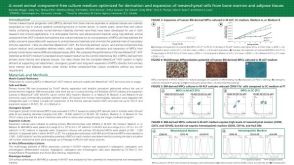 科学海报An Animal Component-Free Chondrogenic Stimulatory Medium for the Efficient Differentiation of Human Mesenchymal Progenitor Cells

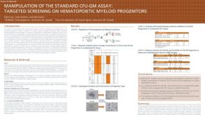 科学海报Manipulation of the Standard CFU-GM Assay Targeted Screening of Hematopoietic Myeloid Progenitors

 沪公网安备31010102008431号