Borchin B et al. (DEC 2013)
Stem Cell Reports 1 6 620--631
Derivation and FACS-Mediated Purification of PAX3+/PAX7+ Skeletal Muscle Precursors from Human Pluripotent Stem Cells
Human pluripotent stem cells (hPSCs) constitute a promising resource for use in cell-based therapies and a valuable in vitro model for studying early human development and disease. Despite significant advancements in the derivation of specific fates from hPSCs,the generation of skeletal muscle remains challenging and is mostly dependent on transgene expression. Here,we describe a method based on the use of a small-molecule GSK3?? inhibitor to derive skeletal muscle from several hPSC lines. We show that early GSK3?? inhibition is sufficient to create the conditions necessary for highly effective derivation of muscle cells. Moreover,we developed a strategy for stringent fluorescence-activated cell sorting-based purification of emerging PAX3+/PAX7+ muscle precursors that are able to differentiate in postsort cultures into mature myocytes. This transgene-free,efficient protocol provides an essential tool for producing myogenic cells for in vivo preclinical studies,in vitro screenings,and disease modeling. ?? 2013 The Authors.
View Publication
产品类型:
产品号#:
05850
05857
05870
05875
85850
85857
85870
85875
产品名:
mTeSR™1
mTeSR™1
D'Alise AM et al. (MAY 2008)
Molecular cancer therapeutics 7 5 1140--9
Reversine, a novel Aurora kinases inhibitor, inhibits colony formation of human acute myeloid leukemia cells.
The demonstration that the small synthetic molecule reversine [2-(4-morpholinoanilino)-N6-cyclohexyladenine] promotes the dedifferentiation of committed cells into multipotent progenitor-type cells has raised hopes on the exploitation of this small chemical tool for the generation of stem cells. Here,we show that reversine causes a failure in cytokinesis and induces polyploidization. These effects of reversine are due to the inhibition of Aurora A and B,two related kinases that are implicated in several aspects of mitosis and that are frequently amplified and overexpressed in human tumors. Reversine inhibits the phosphorylation of histone H3,a direct downstream target of Aurora kinases. Similarly to the Aurora kinase inhibitor VX-680,which has recently entered phase II clinical trials for cancer treatment,reversine inhibited colony formation of leukemic cells from patients with acute myeloid leukemia but was significantly less toxic than VX-680 on cells from healthy donors. The crystal structure of the reversine-Aurora B kinase complex shows that reversine is a novel class of ATP-competitive Aurora kinase inhibitors. Thus,although our studies raise serious doubts on the application of reversine in regenerative medicine,they support the paradigm that reversine might be a useful agent in cancer chemotherapy.
View Publication
产品类型:
产品号#:
72612
72614
产品名:
逆转素(Reversine)
逆转素(Reversine)
Su H et al. (JUL 2013)
Stem Cell Research 11 1 529--539
Transplanted motoneurons derived from human induced pluripotent stem cells form functional connections with target muscle
Induced pluripotent stem cells (iPSCs) hold promise for the treatment of motoneuron diseases because of their distinct features including pluripotency,self-derivation and potential ability to differentiate into motoneurons. However,it is still unknown whether human iPSC-derived motoneurons can functionally innervate target muscles in vivo,which is the definitive sign of successful cell therapy for motoneuron diseases. In the present study,we demonstrated that human iPSCs derived from mesenchymal cells of the umbilical cord possessed a high yield in neural differentiation. Using a chemically-defined in vitro system,human iPSCs efficiently differentiated into motoneurons which displayed typical morphology,expressed specific molecules,and generated repetitive trains of action potentials. When transplanted into the injured musculocutaneous nerve of rats,they survived robustly,extended axons along the nerve,and formed functional connections with the target muscle (biceps brachii),thereby protecting the muscle from atrophy. Our study provides evidence for the first time that human iPSC-derived motoneurons are truly functional not only in vitro but also in vivo,and they have potential for stem cell-based therapies for motoneuron diseases. textcopyright 2013 Elsevier B.V.
View Publication
Cited2 is an essential regulator of adult hematopoietic stem cells.
The regulatory pathways necessary for the maintenance of adult hematopoietic stem cells (HSCs) remain poorly defined. By using loss-of-function approaches,we report a selective and cell-autonomous requirement for the p300/CBP-binding transcriptional coactivator Cited2 in adult HSC maintenance. Conditional deletion of Cited2 in the adult mouse results in loss of HSCs causing multilineage bone marrow failure and increased lethality. In contrast,conditional ablation of Cited2 after lineage specification in lymphoid and myeloid lineages has no impact on the maintenance of these lineages. Additional deletion of Ink4a/Arf (encoding p16(Ink4a) and p19(Arf)) or Trp53 (encoding p53,a downstream target of p19(Arf)) in a Cited2-deficient background restores HSC functionality and rescues mice from bone marrow failure. Furthermore,we show that the critical role of Cited2 in primitive hematopoietic cells is conserved in humans. Taken together,our studies provide genetic evidence that Cited2 selectively maintains adult HSC functions,at least in part,via Ink4a/Arf and Trp53.
View Publication
产品类型:
产品号#:
70008
70008.1
70008.2
70008.3
70008.4
70008.5
70008.6
200-0002
200-0001
200-0000
产品名:
冻存的人脐带血CD34+细胞
冻存的人脐带血CD34+细胞
冻存的人脐带血CD34+细胞
冻存的人脐带血CD34+细胞
冻存的人脐带血CD34+细胞
冻存的人脐带血CD34+细胞
冻存的人脐带血CD34+细胞
冻存的人脐带血CD34+细胞
冻存的人脐带血CD34+细胞
Giassi LJ et al. (AUG 2008)
Experimental biology and medicine (Maywood,N.J.) 233 8 997--1012
Expanded CD34+ human umbilical cord blood cells generate multiple lymphohematopoietic lineages in NOD-scid IL2rgamma(null) mice.
Umbilical cord blood (UCB) is increasingly being used for human hematopoietic stem cell (HSC) transplantation in children but often requires pooling multiple cords to obtain sufficient numbers for transplantation in adults. To overcome this limitation,we have used an ex vivo two-week culture system to expand the number of hematopoietic CD34(+) cells in cord blood. To assess the in vivo function of these expanded CD34(+) cells,cultured human UCB containing 1 x 10(6) CD34(+) cells were transplanted into conditioned NOD-scid IL2rgamma(null) mice. The expanded CD34(+) cells displayed short- and long-term repopulating cell activity. The cultured human cells differentiated into myeloid,B-lymphoid,and erythroid lineages,but not T lymphocytes. Administration of human recombinant TNFalpha to recipient mice immediately prior to transplantation promoted human thymocyte and T-cell development. These T cells proliferated vigorously in response to TCR cross-linking by anti-CD3 antibody. Engrafted TNFalpha-treated mice generated antibodies in response to T-dependent and T-independent immunization,which was enhanced when mice were co-treated with the B cell cytokine BLyS. Ex vivo expanded CD34(+) human UCB cells have the capacity to generate multiple hematopoietic lineages and a functional human immune system upon transplantation into TNFalpha-treated NOD-scid IL2rgamma(null) mice.
View Publication
Gao L et al. (JUL 2016)
Scientific reports 6 29944
Intermittent high oxygen influences the formation of neural retinal tissue from human embryonic stem cells.
The vertebrate retina is a highly multilayered nervous tissue with a large diversity of cellular components. With the development of stem cell technologies,human retinas can be generated in three-dimensional (3-D) culture in vitro. However,understanding the factors modulating key productive processes and the way that they influence development are far from clear. Oxygen,as the most essential element participating in metabolism,is a critical factor regulating organic development. In this study,using 3-D culture of human stem cells,we examined the effect of intermittent high oxygen treatment (40% O2) on the formation and cellular behavior of neural retinas (NR) in the embryonic body (EB). The volume of EB and number of proliferating cells increased significantly under 40% O2 on day 38,50,and 62. Additionally,the ratio of PAX6+ cells within NR was significantly increased. The neural rosettes could only develop with correct apical-basal polarity under 40% O2. In addition,the generation,migration and maturation of retinal ganglion cells were enhanced under 40% O2. All of these results illustrated that 40% O2 strengthened the formation of NR in EB with characteristics similar to the in vivo state,suggesting that the hyperoxic state facilitated the retinal development in vitro.
View Publication
产品类型:
产品号#:
05850
05857
05870
05875
85850
85857
85870
85875
产品名:
mTeSR™1
mTeSR™1
Miyoshi H et al. (JAN 1999)
Science (New York,N.Y.) 283 5402 682--6
Transduction of human CD34+ cells that mediate long-term engraftment of NOD/SCID mice by HIV vectors.
Efficient gene transfer into human hematopoietic stem cells (HSCs) is an important goal in the study of the hematopoietic system as well as for gene therapy of hematopoietic disorders. A lentiviral vector based on the human immunodeficiency virus (HIV) was able to transduce human CD34+ cells capable of stable,long-term reconstitution of nonobese diabetic/severe combined immunodeficient (NOD/SCID) mice. High-efficiency transduction occurred in the absence of cytokine stimulation and resulted in transgene expression in multiple lineages of human hematopoietic cells for up to 22 weeks after transplantation.
View Publication
产品类型:
产品号#:
09500
产品名:
BIT 9500血清替代物
Lu J et al. (MAR 2016)
Stem cells and development 25 9 740--747
Influence of ATM-mediated DNA damage response on genomic variation in human induced pluripotent stem cells.
Genome instability is a potential limitation to the research and therapeutic application of induced pluripotent stem cells (iPSCs). Observed genomic variations reflect the combined activities of DNA damage,cellular DNA damage response (DDR),and selection pressure in culture. To understand the contribution of DDR on the distribution of copy number variations (CNVs) in iPSCs,we mapped CNVs of iPSCs with mutations in the central DDR gene ATM onto genome organization landscapes defined by genome-wide replication timing profiles. We show that following reprogramming the early and late replicating genome is differentially affected by CNVs in ATM deficient iPSCs relative to wild type iPSCs. Specifically,the early replicating regions had increased CNV losses during retroviral reprogramming. This differential CNV distribution was not present after later passage or after episomal reprogramming. Comparison of different reprogramming methods in the setting of defective DNA damage response reveals unique vulnerability of early replicating open chromatin to retroviral vectors.
View Publication
产品类型:
产品号#:
05850
05857
05870
05875
85850
85857
85870
85875
产品名:
mTeSR™1
mTeSR™1
Maetzig T et al. (MAR 2011)
Blood 117 11 3053--64
Polyclonal fluctuation of lentiviral vector-transduced and expanded murine hematopoietic stem cells.
Gene therapy has proven its potential to cure diseases of the hematopoietic system. However,severe adverse events observed in clinical trials have demanded improved gene-transfer conditions. Whereas progress has been made to reduce the genotoxicity of integrating gene vectors,the role of pretransplantation cultivation is less well investigated. We observed that the STIF (stem cell factor [SCF],thrombopoietin [TPO],insulin-like growth factor-2 [IGF-2],and fibroblast growth factor-1 [FGF-1]) cytokine cocktail developed to effectively expand murine hematopoietic stem cells (HSCs) also supports the expansion of leukemia-initiating insertional mutants caused by gammaretroviral gene transfer. We compared 4 protocols to examine the impact of prestimulation and posttransduction culture in STIF in the context of lentiviral gene transfer. Observing 56 transplanted mice for up to 9.5 months,we found consistent engraftment and gene-marking rates after prolonged ex vivo expansion. Although a lentiviral vector with a validated insertional-mutagenic potential was used,longitudinal analysis identifying textgreater 7000 integration sites revealed polyclonal fluctuations,especially in expanded" groups�
View Publication
产品类型:
产品号#:
09600
09650
产品名:
StemSpan™ SFEM
StemSpan™ SFEM
Ratajczak J et al. (AUG 2011)
Leukemia 25 8 1278--85
Hematopoietic differentiation of umbilical cord blood-derived very small embryonic/epiblast-like stem cells.
A population of CD133(+)Lin(-)CD45(-) very small embryonic/epiblast-like stem cells (VSELs) has been purified by multiparameter sorting from umbilical cord blood (UCB). To speed up isolation of these cells,we employed anti-CD133-conjugated paramagnetic beads followed by staining with Aldefluor to detect aldehyde dehydrogenase (ALDH) activity; we subsequently sorted CD45(-)/GlyA(-)/CD133(+)/ALDH(high) and CD45(-)/GlyA(-)/CD133(+)/ALDH(low) cells,which are enriched for VSELs,and CD45(+)/GlyA /CD133(+)/ALDH(high) and CD45(+)/GlyA(-)/CD133(+)/ALDH(low) cells,which are enriched for hematopoietic stem/progenitor cells (HSPCs). Although freshly isolated CD45(-) VSELs did not grow hematopoietic colonies,the same cells,when activated/expanded over OP9 stromal support,acquired hematopoietic potential and grew colonies composed of CD45(+) hematopoietic cells in methylcellulose cultures. We also observed that CD45(-)/GlyA(-)/CD133(+)/ALDH(high) VSELs grew colonies earlier than CD45(-)/GlyA(-)/CD133(+)/ALDH(low) VSELs,which suggests that the latter cells need more time to acquire hematopoietic commitment. In support of this possibility,real-time polymerase chain reaction analysis confirmed that,whereas freshly isolated CD45(-)/GlyA(-)/CD133(+)/ALDH(high) VSELs express more hematopoietic transcripts (for example,c-myb),CD45(-)/GlyA(-)/CD133(+)/ALDH(low) VSELs exhibit higher levels of pluripotent stem cell markers (for example,Oct-4). More importantly,hematopoietic cells derived from VSELs that were co-cultured over OP9 support were able to establish human lympho-hematopoietic chimerism in lethally irradiated non-obese diabetic/severe combined immunodeficiency mice 4-6 weeks after transplantation. Overall,our data suggest that UCB-VSELs correspond to the most primitive population of HSPCs in UCB.
View Publication

 EasySep™小鼠TIL(CD45)正选试剂盒


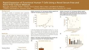 科学海报Rapid Expansion of Functional Human T Cells Using a Novel Serum-Free and Xeno-Free Culture Medium

 沪公网安备31010102008431号