
 EasySep™小鼠TIL(CD45)正选试剂盒


产品号 #05507_C
用于小鼠MSCs、ADSCs和MEFs体外向脂肪细胞的分化
若您需要咨询产品或有任何技术问题,请通过官方电话 400 885 9050 或邮箱 info.cn@stemcell.com 与我们联系。
用于小鼠MSCs、ADSCs和MEFs体外向脂肪细胞的分化
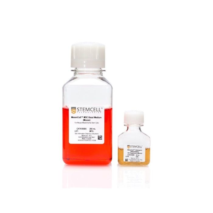
MesenCult™ 脂肪分化试剂盒 (小鼠) 专门用于小鼠间充质干细胞或祖细胞(MSCs),小鼠脂肪组织来源的MSCs (ADSCs)和小鼠胚胎成纤维细胞(MEFs)体外分化成脂肪生成谱系的细胞。
注意:MesenCult™ 脂肪分化试剂盒 (小鼠)必须补充 L-谷氨酰胺(产品号 #07100)。
分类
专用培养基
细胞类型
间充质干/祖细胞
种属
小鼠
应用
分化
品牌
MesenCult
研究领域
药物发现和毒性检测,干细胞生物学
请在《产品说明书》中查找相关支持信息和使用说明,或浏览下方更多实验方案。
本产品专为以下研究领域设计,适用于工作流程中的高亮阶段。探索这些工作流程,了解更多我们为各研究领域提供的其他配套产品。
| 物种 | 小鼠 |
|---|
小鼠间充质干细胞和胚胎成纤维细胞分化为成骨细胞的完全培养基
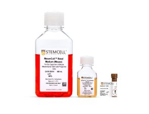
用于培养小鼠MSCs和MEFs
<p>对来源于小鼠致密骨或其他组织单细胞悬液的未标记间充质干细胞进行免疫磁珠分离</p>
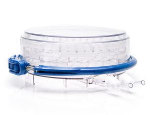
培养室为组织培养提供缺氧环境
在线联系


 沪公网安备31010102008431号