若您需要咨询产品或有任何技术问题,请通过官方电话 400 885 9050 或邮箱 info.cn@stemcell.com 与我们联系

MesenCult™ 增殖试剂盒(人)

检测CFU-F和人间充质干细胞扩增的培养基

产品号 #(选择产品)

产品号 #05411_C

检测CFU-F和人间充质干细胞扩增的培养基

产品组分包括

  • MesenCult™ MSC 基础培养基(人),450 mL(产品号 #05401);
  • MesenCult™ MSC 刺激补充剂(人),50 mL(产品号 #05402)

总览

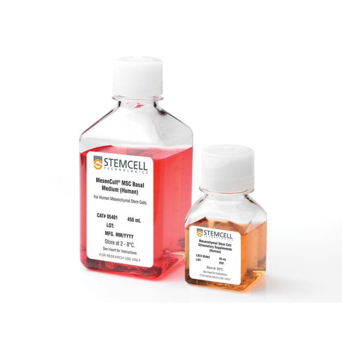
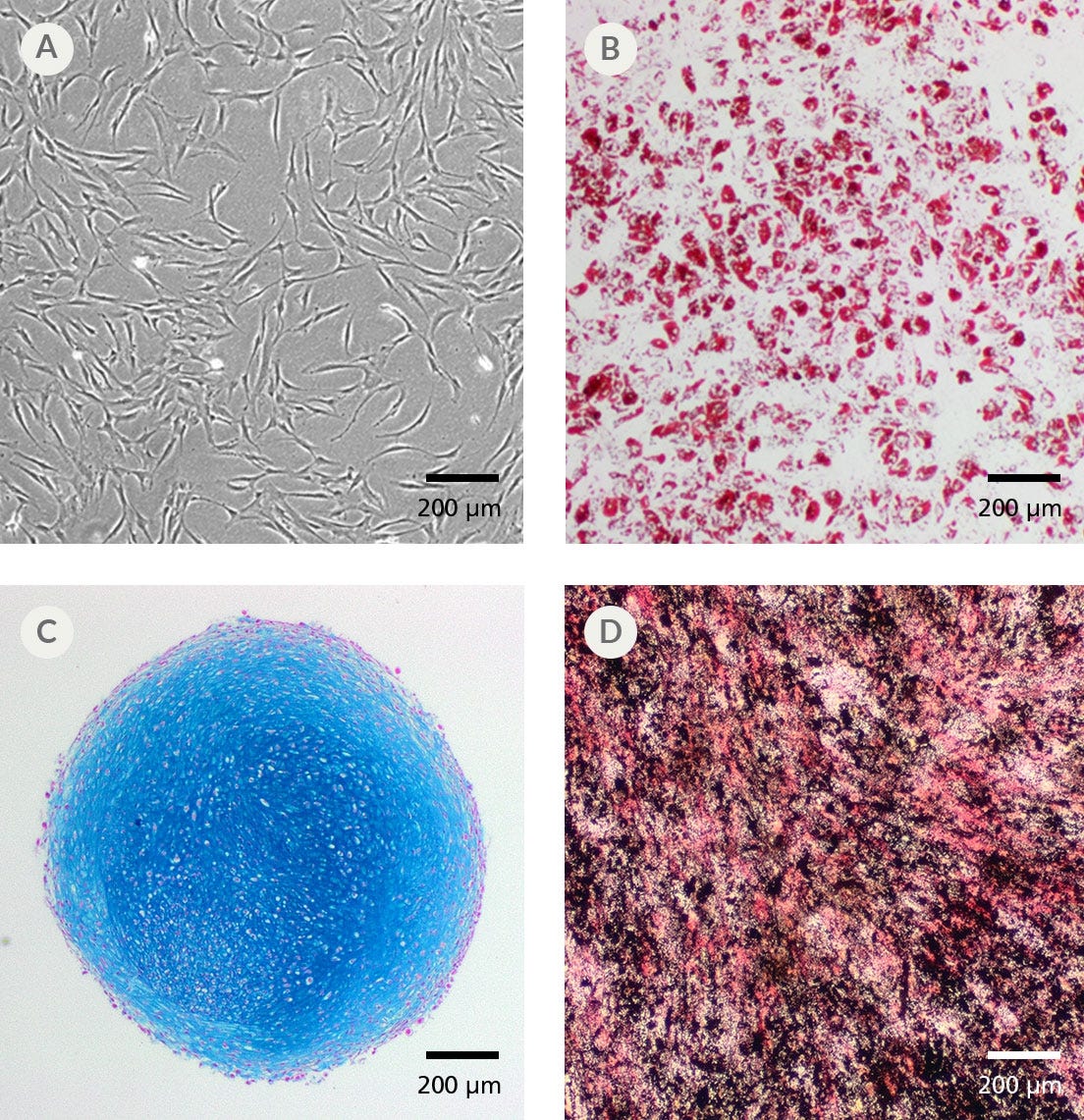
MesenCult™ 增殖试剂盒(人)是一种标准化的含血清培养基,用于培养人间充质干细胞(MSCs)。MesenCult™ 增殖试剂盒(人)已经过优化,可用于人MSCs的体外扩增以及集落形成单位-成纤维细胞(CFU-F)的检测和计数。MesenCult™ 增殖试剂盒(人)包含 MesenCult™ MSC基础培养基(人;450 mL)和 MesenCult™ MSC刺激补充剂(人;50 mL)两个组分。

亚型
专用培养基
 
细胞类型
间充质干/祖细胞
 
种属

 
应用
细胞培养,克隆筛选,扩增
 
品牌
MesenCult
 
研究领域
干细胞生物学
 

实验数据

Figure 1. Human Mesenchymal Stem and Progenitor cells (MSCs) Derived and Culture-Expanded Using the MesenCult™ Proliferation Kit

(A) Human bone marrow mesenchymal stem and progenitor cells (BM MSCs) were derived and culture-expanded for 8 passages in complete MesenCult™ Proliferation Medium (n = 6) and two commercially available media (Commercial Medium 1 and Commercial Medium 2; n = 3). (B) The average doubling time in days over 8 passages of human BM MSCs culture-expanded in complete MesenCult™ Proliferation Medium, and Commercial Medium 1 and 2. Vertical lines indicate standard error of the mean.

Figure 2. Human MSCs Culture-Expanded in Complete MesenCult™ Proliferation Medium Retain Strong Multi-Lineage Differentiation Potential

(A) Human BM-derived MSCs cultured in complete MesenCult™ Proliferation Medium differentiate into (B) adipocytes (Oil Red O staining), (C) chondrocytes (Alcian Blue staining) and (D) osteoblasts (alkaline phosphatase and von Kossa staining).

Figure 3. Flow Cytometric Analysis of Culture-Expanded Human MSCs in Complete MesenCult™ Proliferation Medium

Human BM-derived MSCs from passage 5 were stained for mesenchymal cell surface markers CD73, CD90 and CD105, and the hematopoietic markers CD34 and CD45 (shown in grey shaded areas). BM-derived MSCs are positive for MSC markers and negative for hematopoietic markers. High expression of the endothelial marker, CD146, was also observed in BM-derived MSCs (shown in grey shaded areas). Isotype control is shown by red line.

产品说明书及文档

请在《产品说明书》中查找相关支持信息和使用说明,或浏览下方更多实验方案。

Document Type
Product Name
Catalog #
Lot #
Language
Catalog #
05411
Lot #
All
Language
English
Document Type
Safety Data Sheet 1
Catalog #
05411
Lot #
All
Language
English
Document Type
Safety Data Sheet 2
Catalog #
05411
Lot #
All
Language
English

应用领域

本产品专为以下研究领域设计,适用于工作流程中的高亮阶段。探索这些工作流程,了解更多我们为各研究领域提供的其他配套产品。

相关材料与文献

技术资料 (9)

文献 (39)

Thymosin $\beta$4-Enhancing Therapeutic Efficacy of Human Adipose-Derived Stem Cells in Mouse Ischemic Hindlimb Model. J.-H. Kim et al. International journal of molecular sciences 2020 mar

Abstract

Thymosin $\beta$4 (T$\beta$4) is a G-actin sequestering protein that contributes to diverse cellular activities, such as migration and angiogenesis. In this study, the beneficial effects of combined cell therapy with T$\beta$4 and human adipose-derived stem cells (hASCs) in a mouse ischemic hindlimb model were investigated. We observed that exogenous treatment with T$\beta$4 enhanced endogenous TMSB4X mRNA expression and promoted morphological changes (increased cell length) in hASCs. Interestingly, T$\beta$4 induced the active state of hASCs by up-regulating intracellular signaling pathways including the PI3K/AKT/mTOR and MAPK/ERK pathways. Treatment with T$\beta$4 significantly increased cell migration and sprouting from microbeads. Moreover, additional treatment with T$\beta$4 promoted the endothelial differentiation potential of hASCs by up-regulating various angiogenic genes. To evaluate the in vivo effects of the T$\beta$4-hASCs combination on vessel recruitment, dorsal window chambers were transplanted, and the co-treated mice were found to have a significantly increased number of microvessel branches. Transplantation of hASCs in combination with T$\beta$4 was found to improve blood flow and attenuate limb or foot loss post-ischemia compared to transplantation with hASCs alone. Taken together, the therapeutic application of hASCs combined with T$\beta$4 could be effective in enhancing endothelial differentiation and vascularization for treating hindlimb ischemia.
New approach to isolate mesenchymal stem cell (MSC) from human umbilical cord blood. Hussain I et al. Cell biology international 2012 JUL

Abstract

HUCB (human umbilical cord blood) has been frequently used in clinical allogeneic HSC (haemopoietic stem cell) transplant. However, HUCB is poorly recognized as a rich source of MSC (mesenchymal stem cell). The aim of this study has been to establish a new method for isolating large number of MSC from HUCB to recognize it as a good source of MSC. HUCB samples were collected from women following their elective caesarean section. The new method (Clot Spot method) was carried out by explanting HUCB samples in mesencult complete medium and maintained in 37°C, in a 5% CO2 and air incubator. MSC presence was established by quantitative and qualitative immunophenotyping of cells and using FITC attached to MSC phenotypic markers (CD29, CD73, CD44 and CD105). Haematopoietic antibodies (CD34 and CD45) were used as negative control. MSC differentiation was examined in neurogenic and adipogenic media. Immunocytochemistry was carried out for the embryonic markers: SOX2 (sex determining region Y-box 2), OLIG-4 (oligodendrocyte-4) and FABP-4 (fatty acid binding protein-4). The new method was compared with the conventional Rosset Sep method. MSC cultures using the Clot Spot method showed 3-fold increase in proliferation rate compared with conventional method. Also, the cells showed high expression of MSC markers CD29, CD73, CD44 and CD105, but lacked the expression of specific HSC markers (CD34 and CD45). The isolated MSC showed some differentiation by expressing the neurogenic (SOX2 and Olig4) and adipogenic (FABP-4) markers respectively. In conclusion, HUCB is a good source of MSC using this new technique.
Chondrogenesis by chemotactic homing of synovium, bone marrow, and adipose stem cells in vitro. Mendelson A et al. FASEB journal : official publication of the Federation of American Societies for Experimental Biology 2011 OCT

Abstract

Cell transplantation has been well explored for cartilage regeneration. We recently showed that the entire articular surface of a synovial joint can regenerate by endogenous cell homing and without cell transplantation. However, the sources of endogenous cells that regenerate articular cartilage remain elusive. Here, we studied whether cytokines not only chemotactically recruit adipose stem cells (ASCs), mesenchymal stem cells (MSCs), and synovium stem cells (SSCs) but also induce chondrogenesis of the recruited cells. Recombinant human transforming growth factor-β3 (TGF-β3; 100 ng) and/or recombinant human stromal derived factor-1β (SDF-1β; 100 ng) was control released into an acellular collagen sponge cube with underlying ASCs, MSCs, or SSCs in monolayer culture. Although all cell types randomly migrated into the acellular collagen sponge cube, TGF-β3 and/or SDF-1β recruited significantly more cells than the cytokine-free control group. In 6 wk, TGF-β3 alone recruited substantial numbers of ASCs (558±65) and MSCs (302±52), whereas codelivery of TGF-β3 and SDF-1β was particularly chemotactic to SSCs (400±120). Proliferation of the recruited cells accounted for some, but far from all, of the observed cellularity. TGF-β3 and SDF-1β codelivery induced significantly higher aggrecan gene expression than the cytokine-free group for ASCs, MSCs, and SSCs. Type II collagen gene expression was also significantly higher for ASCs and SSCs by SDF-1 and TGF-β3 codelivery. Remarkably, the expression of aggrecan and type II collagen was detected among all cell types. Thus, homing of multiple stem/progenitor cell populations may potentially serve as an alternative or adjunctive approach to cell transplantation for cartilage regeneration.

更多信息

更多信息
种属 Human
质量保证:

产品仅供研究使用,不用于针对人或动物的诊断或治疗。 欲获悉更多关于STEMCELL的质控信息,请访问 STEMCELL.CN/COMPLIANCE.
Copyright © 2025 by STEMCELL Technologies. All rights reserved.