Kordon EC and Smith GH (MAY 1998)
Development (Cambridge,England) 125 10 1921--30
An entire functional mammary gland may comprise the progeny from a single cell.
Any epithelial portion of a normal mouse mammary gland can reproduce an entire functional gland when transplanted into an epithelium-free mammary fat pad. Mouse mammary hyperplasias and tumors are clonal dominant populations and probably represent the progeny of a single transformed cell. Our study provides evidence that single multipotent stem cells positioned throughout the mature fully developed mammary gland have the capacity to produce sufficient differentiated progeny to recapitulate an entire functional gland. Our evidence also demonstrates that these stem cells are self-renewing and are found with undiminished capacities in the newly regenerated gland. We have taken advantage of an experimental model where mouse mammary tumor virus infects mammary epithelial cells and inserts a deoxyribonucleic acid copy(ies) of its genome during replication. The insertions occur randomly within the somatic genome. CzechII mice have no endogenous nucleic acid sequence homology with mouse mammary tumor virus; therefore all viral insertions may be detected by Southern analysis provided a sufficient number of cells contain a specific insertional event. Transplantation of random fragments of infected CzechII mammary gland produced clonal-dominant epithelial populations in epithelium-free mammary fat pads. Serial transplantation of pieces of the clonally derived outgrowths produced second generation glands possessing the same viral insertion sites providing evidence for self-renewal of the original stem cell. Limiting dilution studies with cell cultures derived from third generation clonal outgrowths demonstrated that three multipotent but distinct mammary epithelial progenitors were present in clonally derived mammary epithelial populations. Estimation of the potential number of multipotent epithelial cells that may be evolved from an individual mammary-specific stem cell by self-renewal is in the order of 10(12)-10(13). Therefore,one stem cell might easily account for the renewal of mammary epithelium over several transplant generations.
View Publication
文献
Smith GH (JAN 1996)
Breast cancer research and treatment 39 1 21--31
Experimental mammary epithelial morphogenesis in an in vivo model: evidence for distinct cellular progenitors of the ductal and lobular phenotype.
An in vivo transplantation system has been used to evaluate the developmental capacities of specific mouse mammary epithelial cell populations. Specifically,mouse mammary epithelial cells with distinctly limited developmental potentials have been identified using this procedure. Two distinct epithelial cell progenitors have been identified by experiments designed to determine whether basal lobular and ductal phenotypes could develop independently under conditions imposed by a limiting dilution. The prediction that these separate epithelial progenitors must exist was based upon the results from transplantation experiments carried out in epithelium-divested mammary fat pads of syngeneic mice with mammary epithelium from two different transgenic mouse models. The results presented here demonstrate the following points: 1) lobular,i.e. secretory,progenitor cells are present as distinct entities among the mammary epithelial cells found in immature virgin female mice; 2) similarly,ductal epithelial progenitors are present within the same population; 3) lobular progenitors are present in greater numbers,although both cell populations are extremely small; 4) as expected,some inocula produce outgrowths with simultaneous development of both lobular and ductal phenotypes--it is not known whether this indicates cooperative interaction between the two epithelial progenitors or signals the presence of a third progenitor type capable of producing both ductular and lobular committed daughters; 5) these findings have important consequences in the design of experiments aimed at testing the effects of known and putative mammary oncogenes and tumor suppressor genes,using techniques which include cellular transformation in vitro followed by in vivo cultivation and evaluation.
View Publication
文献
Kumar A et al. (JAN 2012)
Breast cancer research : BCR 14 1 R4
Evidence that GTP-binding domain but not catalytic domain of transglutaminase 2 is essential for epithelial-to-mesenchymal transition in mammary epithelial cells.
INTRODUCTION: The expression of proinflammatory protein tissue transglutaminase 2 (TG2) is frequently upregulated in multiple cancer cell types. However,the exact role of TG2 in cancer cells is not well-understood. We recently initiated studies to determine the significance of TG2 in cancer cells and observed that sustained expression of TG2 resulted in epithelial-to-mesenchymal transition (EMT) and promoted cancer stem cell (CSC) traits in mammary epithelial cells. These results suggested that TG2 could serve as a promising therapeutic target for overcoming chemoresistance and inhibiting metastatic spread of cancer cells. METHODS: Using various mutant constructs,we analyzed the activity of TG2 that is essential for promoting the EMT-CSC phenotype. RESULTS: Our results suggest that catalytically inactive TG2 (TG2-C277S) is as effective as wild-type TG2 (TG2-WT) in inducing the EMT-CSC in mammary epithelial cells. In contrast,overexpression of a GTP-binding-deficient mutant (TG2-R580A) was completely incompetent in this regard. Moreover,TG2-dependent activation of the proinflammatory transcription factor NF-κB is deemed essential for promoting the EMT-CSC phenotype in mammary epithelial cells. CONCLUSIONS: Our results suggest that the transamidation activity of TG2 is not essential for promoting its oncogenic functions and provide a strong rationale for developing small-molecule inhibitors to block GTP-binding pockets of TG2. Such inhibitors may have great potential for inhibiting the TG2-regulated pathways,reversing drug resistance and inhibiting the metastasis of cancer cells.
View Publication
文献
Liu C et al. (MAY 2012)
Molecular biology reports 39 5 5875--81
Co-expression of Oct-4 and Nestin in human breast cancers.
The aim is to investigate the clinical implications of the Oct-4 and Nestin protein in human breast cancers. A total of 346 cases including 26 fresh and 320 paraffin-embedded tumor tissues were selected for characterizing the frequency of CD44(+)CD24(-) tumor cells by flow cytometry and the differential expression of the stem cell-related genes between CD44(+)CD24(-) and non-CD44(+)CD24(-) tumor cells was analyzed by PCR Array and immunofluorescence. In comparison with the non-CD44(+)CD24(-) tumor cells,the CD44(+)CD24(-),particularly for those with high percentage of Oct-4(+) and Nestin(+),tumor cells had higher tumorigenicity by forming mammospheres in vitro. More importantly,42 (13.125%) out of 320 tumor tissues were positive for Oct-4 and Nestin staining. Universal analysis and multivariate analysis revealed that the expression of Oct-4 and Nestin was associated significantly with younger age,pathogenic degrees,lymph node metastasis and triple-negative breast cancer independently (P textless 0.05) as well as shorter survival (P = 0.001). Oct-4 and Nestin were important regulators of the development of breast cancer,and Oct-4 and Nestin may be used as predictors for the prognosis of breast cancers.
View Publication
文献
Wu H et al. (SEP 2011)
Journal of breast cancer 14 3 175--80
Can CD44+/CD24- Tumor Cells Be Used to Determine the Extent of Breast Cancer Invasion Following Neoadjuvant Chemotherapy?
PURPOSE: To investigate the distribution of CD44(+)/CD24(-) cells in breast cancers in relation to tumor size before and after the administration of neoadjuvant chemotherapy. METHODS: CD44(+)/CD24(-) tumor cells obtained from breast cancer specimens were characterized in vivo and in vitro using tumor formation assays and mammosphere generation assays,respectively. The distribution of CD44+/CD24- tumor cells in 78 breast cancer specimens following administration of neoadjuvant chemotherapy was also evaluated using immunofluorescence assays,and this distribution was compared with the extent of tumor invasion predicted by Response Evaluation Criteria in Solid Tumours (RECIST). RESULTS: In 27/78 cases,complete remission (CR) was identified using RECIST. However,18 of these CR cases were associated with a scattered distribution of tumor stem cells in the outline of the original tumor prior to neoadjuvant chemotherapy. After neoadjuvant chemotherapy,24 cases involved cancer cells that were confined to the tumor outline,and 21 cases had tumor cells or tumor stem cells overlapping the tumor outline. In addition,there were 6 patients who were insensitive to chemotherapy,and in these cases,both cancer cells and stem cells were detected outside the contours of the tumor volume imaged prior to chemotherapy. CONCLUSION: CD44+/CD24- tumor cells may be an additional parameter to evaluate when determining the extent of breast cancer invasion.
View Publication
文献
Marcato P et al. (MAY 2011)
Cell cycle (Georgetown,Tex.) 10 9 1378--84
Aldehyde dehydrogenase: its role as a cancer stem cell marker comes down to the specific isoform.
Recent evidence suggests that enhanced aldehyde dehydrogenase (ALDH) activity is a hallmark of cancer stem cells (CSC) measurable by the aldefluor assay. ALDH1A1,one of 19 ALDH isoforms expressed in humans,was generally believed to be responsible for the ALDH activity of CSCs. More recently,experiments with murine hematopoietic stem cells,murine progenitor pancreatic cells,and human breast CSCs indicate that other ALDH isoforms,particularly ALDH1A3,significantly contribute to aldefluor positivity,which may be tissue and cancer specific. Therefore,potential prognostic application involving the use of CSC prevalence in tumor tissue to predict patient outcome requires the identification and quantification of specific ALDH isoforms. Herein we review the suggested roles of ALDH in CSC biology and the immunohistological studies testing the potential application of ALDH isoforms as novel cancer prognostic indicators.
View Publication
文献
De Giorgi U et al. (MAY 2011)
Cancer biology & therapy 11 9 812--5
Mesenchymal stem cells expressing GD2 and CD271 correlate with breast cancer-initiating cells in bone marrow.
Purpose: The bone marrow microenvironment is considered a critical component in the dissemination and fate of cancer cells in the metastatic process. We explored the possible correlation between bone marrow mesenchymal stem cells (BM-MSC) and disseminated breast cancer-initiating cells (BCIC) in primary breast cancer patients. Experimental design: Bone marrow mononuclear cells (BM-MNC) were collected at the time of primary surgery in 12 breast cancer patients. BM-MNC was immunophenotyped and BCIC was defined as epithelial cells (CD326+CD45-) with a stem-like" phenotype (CD44+CD24low/-�
View Publication
文献
Liu S et al. (JAN 2011)
Cancer research 71 2 614--24
Breast cancer stem cells are regulated by mesenchymal stem cells through cytokine networks.
We have used in vitro and mouse xenograft models to examine the interaction between breast cancer stem cells (CSC) and bone marrow-derived mesenchymal stem cells (MSC). We show that both of these cell populations are organized in a cellular hierarchy in which primitive aldehyde dehydrogenase expressing mesenchymal cells regulate breast CSCs through cytokine loops involving IL6 and CXCL7. In NOD/SCID mice,labeled MSCs introduced into the tibia traffic to sites of growing breast tumor xenografts where they accelerated tumor growth by increasing the breast CSC population. With immunochemistry,we identified MSC-CSC niches in these tumor xenografts as well as in frozen sections from primary human breast cancers. Bone marrow-derived MSCs may accelerate human breast tumor growth by generating cytokine networks that regulate the CSC population.
View Publication
文献
Ma I and Allan AL (JUN 2011)
Stem cell reviews 7 2 292--306
The role of human aldehyde dehydrogenase in normal and cancer stem cells.
Normal stem cells and cancer stem cells (CSCs) share similar properties,in that both have the capacity to self-renew and differentiate into multiple cell types. In both the normal stem cell and cancer stem cell fields,there has been a great need for a universal marker that can effectively identify and isolate these rare populations of cells in order to characterize them and use this information for research and therapeutic purposes. Currently,it would appear that certain isoenzymes of the aldehyde dehydrogenase (ALDH) superfamily may be able to fulfill this role as a marker for both normal and cancer stem cells. ALDH has been identified as an important enzyme in the protection of normal hematopoietic stem cells,and is now also widely used as a marker to identify and isolate various types of normal stem cells and CSCs. In addition,emerging evidence suggests that ALDH1 is not only a marker for stem cells,but may also play important functional roles related to self-protection,differentiation,and expansion. This comprehensive review discusses the role that ALDH plays in normal stem cells and CSCs,with focus on ALDH1 and ALDH3A1. Discrepancies in the functional themes between cell types and future perspectives for therapeutic applications will also be discussed.
View Publication
文献
Alison MR et al. (DEC 2010)
The Journal of pathology 222 4 335--44
Finding cancer stem cells: are aldehyde dehydrogenases fit for purpose?
Despite many years of intensive effort,there is surprisingly little consensus on the most suitable markers with which to locate and isolate stem cells from adult tissues. By comparison,the study of cancer stem cells is still in its infancy; so,unsurprisingly,there is great uncertainty as to the identity of these cells. Stem cell markers can be broadly categorized into molecular determinants of self-renewal,clonogenicity,multipotentiality,adherence to the niche,and longevity. This review assesses the utility of recognizing cancer stem cells by virtue of high expression of aldehyde dehydrogenases (ALDHs),probably significant determinants of cell survival through their ability to detoxify many potentially cytotoxic molecules,and contributing to drug resistance. Antibodies are available against the ALDH enzyme family,but the vast majority of studies have used cell sorting techniques to enrich for cells expressing these enzymes. Live cells expressing high ALDH activity are usually identified by the ALDEFLUOR kit and sorted by fluorescence activated cell sorting (FACS). For many human tumours,but notably breast cancer,cell selection based upon ALDH activity appears to be a useful marker for enriching for cells with tumour-initiating activity (presumed cancer stem cells) in immunodeficient mice,and indeed the frequency of so-called ALDH(bri) cells in many tumours can be an independent prognostic indicator.
View Publication

 EasySep™小鼠TIL(CD45)正选试剂盒


 Mini ReviewMammary Stem Cells and Breast Cancer
 文献
文献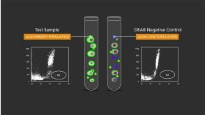

 沪公网安备31010102008431号