Enhanced erythropoiesis in Hfe-KO mice indicates a role for Hfe in the modulation of erythroid iron homeostasis.
In hereditary hemochromatosis,mutations in HFE lead to iron overload through abnormally low levels of hepcidin. In addition,HFE potentially modulates cellular iron uptake by interacting with transferrin receptor,a crucial protein during erythropoiesis. However,the role of HFE in this process was never explored. We hypothesize that HFE modulates erythropoiesis by affecting dietary iron absorption and erythroid iron intake. To investigate this,we used Hfe-KO mice in conditions of altered dietary iron and erythropoiesis. We show that Hfe-KO mice can overcome phlebotomy-induced anemia more rapidly than wild-type mice (even when iron loaded). Second,we evaluated mice combining the hemochromatosis and β-thalassemia phenotypes. Our results suggest that lack of Hfe is advantageous in conditions of increased erythropoietic activity because of augmented iron mobilization driven by deficient hepcidin response. Lastly,we demonstrate that Hfe is expressed in erythroid cells and impairs iron uptake,whereas its absence exclusively from the hematopoietic compartment is sufficient to accelerate recovery from phlebotomy. In summary,we demonstrate that Hfe influences erythropoiesis by 2 distinct mechanisms: limiting hepcidin expression under conditions of simultaneous iron overload and stress erythropoiesis,and impairing transferrin-bound iron uptake by erythroid cells. Moreover,our results provide novel suggestions to improve the treatment of hemochromatosis.
View Publication
产品号#:
03334
产品名:
MethoCult™ M3334
Lam RS et al. ( 2017)
PloS one 12 1 e0169506
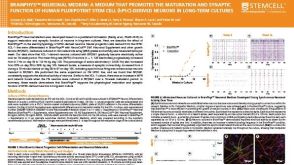
Functional Maturation of Human Stem Cell-Derived Neurons in Long-Term Cultures.
Differentiated neurons can be rapidly acquired,within days,by inducing stem cells to express neurogenic transcription factors. We developed a protocol to maintain long-term cultures of human neurons,called iNGNs,which are obtained by inducing Neurogenin-1 and Neurogenin-2 expression in induced pluripotent stem cells. We followed the functional development of iNGNs over months and they showed many hallmark properties for neuronal maturation,including robust electrical and synaptic activity. Using iNGNs expressing a variant of channelrhodopsin-2,called CatCh,we could control iNGN activity with blue light stimulation. In combination with optogenetic tools,iNGNs offer opportunities for studies that require precise spatial and temporal resolution. iNGNs developed spontaneous network activity,and these networks had excitatory glutamatergic synapses,which we characterized with single-cell synaptic recordings. AMPA glutamatergic receptor activity was especially dominant in postsynaptic recordings,whereas NMDA glutamatergic receptor activity was absent from postsynaptic recordings but present in extrasynaptic recordings. Our results on long-term cultures of iNGNs could help in future studies elucidating mechanisms of human synaptogenesis and neurotransmission,along with the ability to scale-up the size of the cultures.
View Publication
产品号#:
05854
05855
85850
85857
85870
85875
产品名:
mFreSR™
mFreSR™
mTeSR™1
mTeSR™1
Wei Y et al. (MAR 2017)
Placenta 51 28--37
Generation of trophoblast-like cells from the amnion in vitro: A novel cellular model for trophoblast development.
Despite the high incidence of trophoblast-related diseases,the molecular mechanism of inadequate early trophoblast development is still unclear due to the lack of an appropriate cellular model in vitro. In the present study,we reprogrammed the amniotic cells to be induced pluripotent stem cells (iPSCs) via a non-virus and non-integrated method and subsequently differentiated them into trophoblast-like cells by a modified BMP4 strategy in E6 medium. Compared with the previously studied trophoblast-like cells from ESCs,the iPSCs derived trophoblast-like cells behave similarly in terms of gene expression profiles and biofunctions. Also we confirmed the differentiating tendency from iPSCs to be syncytiotrophoblasts-like cells might be caused by inappropriate differentiating oxygen condition. Additionally,we preliminarily indicated in vitro artificial" differentiation of iPSCs also undergoing a possible trophoblastic stem cell stage as witnessed in vivo. In conclusion we provided an in vitro cellular model to study early trophoblast development for specific individual by using the feasible amnion.
View Publication
产品号#:
85850
85857
85870
85875
产品名:
mTeSR™1
mTeSR™1
Wills QF et al. (JAN 2017)
BMC genomics 18 1 53
The nature and nurture of cell heterogeneity: accounting for macrophage gene-environment interactions with single-cell RNA-Seq.
BACKGROUND Single-cell RNA-Seq can be a valuable and unbiased tool to dissect cellular heterogeneity,despite the transcriptome's limitations in describing higher functional phenotypes and protein events. Perhaps the most important shortfall with transcriptomic 'snapshots' of cell populations is that they risk being descriptive,only cataloging heterogeneity at one point in time,and without microenvironmental context. Studying the genetic ('nature') and environmental ('nurture') modifiers of heterogeneity,and how cell population dynamics unfold over time in response to these modifiers is key when studying highly plastic cells such as macrophages. RESULTS We introduce the programmable Polaris microfluidic lab-on-chip for single-cell sequencing,which performs live-cell imaging while controlling for the culture microenvironment of each cell. Using gene-edited macrophages we demonstrate how previously unappreciated knockout effects of SAMHD1,such as an altered oxidative stress response,have a large paracrine signaling component. Furthermore,we demonstrate single-cell pathway enrichments for cell cycle arrest and APOBEC3G degradation,both associated with the oxidative stress response and altered proteostasis. Interestingly,SAMHD1 and APOBEC3G are both HIV-1 inhibitors ('restriction factors'),with no known co-regulation. CONCLUSION As single-cell methods continue to mature,so will the ability to move beyond simple 'snapshots' of cell populations towards studying the determinants of population dynamics. By combining single-cell culture,live-cell imaging,and single-cell sequencing,we have demonstrated the ability to study cell phenotypes and microenvironmental influences. It's these microenvironmental components - ignored by standard single-cell workflows - that likely determine how macrophages,for example,react to inflammation and form treatment resistant HIV reservoirs.
View Publication
产品号#:
85850
85857
85870
85875
产品名:
mTeSR™1
mTeSR™1
Deng Y et al. (FEB 2017)
Biomacromolecules 18 2 587--598
Peptide-Decorated Nanofibrous Niche Augments In Vitro Directed Osteogenic Conversion of Human Pluripotent Stem Cells.
Realization of clinical potential of human pluripotent stem cells (hPSCs) in bone regenerative medicine requires development of simple and safe biomaterials for expansion of hPSCs followed by directing their lineage commitment to osteoblasts. In the present study,a chemically defined peptide-decorated polycaprolactone (PCL) nanofibrous microenvironment was prepared through electrospinning technology and subsequent conjugation with vitronectin peptide to promote the culture and osteogenic potential of hPSCs in vitro. The results indicated that hPSCs successfully proliferated and maintained their pluripotency on the biointerface of peptide-conjugated nanofibers without Matrigel under defined conditions. Moreover,the prepared niche exhibited an appealing ability in promoting directed differentiation of hPSCs to osteoblastic phenotype without embryoid body formation step,determined from the cell morphological alteration,alkaline phosphate activity,and osteogenesis-related gene expression,as well as protein production. Such well-defined,xeno-free,and safe nanofiber scaffolds that allow the survival and facilitate osteo-differentiation of hPSCs provide a novel platform for hPSCs differentiation via cell-nanofiber interplay,and possess great value in accelerating the translational perspectives of hPSCs in bone tissue engineering.
View Publication
Maillet A et al. ( 2016)
Scientific reports 6 April 25333
Modeling Doxorubicin-Induced Cardiotoxicity in Human Pluripotent Stem Cell Derived-Cardiomyocytes.
Doxorubicin is a highly efficacious anti-cancer drug but causes cardiotoxicity in many patients. The mechanisms of doxorubicin-induced cardiotoxicity (DIC) remain incompletely understood. We investigated the characteristics and molecular mechanisms of DIC in human pluripotent stem cell-derived cardiomyocytes (hPSC-CMs). We found that doxorubicin causes dose-dependent increases in apoptotic and necrotic cell death,reactive oxygen species production,mitochondrial dysfunction and increased intracellular calcium concentration. We characterized genome-wide changes in gene expression caused by doxorubicin using RNA-seq,as well as electrophysiological abnormalities caused by doxorubicin with multi-electrode array technology. Finally,we show that CRISPR-Cas9-mediated disruption of TOP2B,a gene implicated in DIC in mouse studies,significantly reduces the sensitivity of hPSC-CMs to doxorubicin-induced double stranded DNA breaks and cell death. These data establish a human cellular model of DIC that recapitulates many of the cardinal features of this adverse drug reaction and could enable screening for protective agents against DIC as well as assessment of genetic variants involved in doxorubicin response.
View Publication
产品号#:
05850
05857
05870
05875
85850
85857
85870
85875
产品名:
mTeSR™1
mTeSR™1
Galat V et al. (MAY 2016)
Stem cells and development 25 14 1060--1072
Transgene Reactivation in Induced Pluripotent Stem Cell Derivatives and Reversion to Pluripotency of Induced Pluripotent Stem Cell-Derived Mesenchymal Cells.
Induced pluripotent stem cells (iPSCs) have enormous potential in regenerative medicine and disease modeling. It is now felt that clinical trials should be performed with iPSCs derived with non-integrative constructs. Numerous studies,however,including those describing disease models,are still being published using cells derived from iPSCs generated with integrative constructs. Our experimental work presents the first evidence of spontaneous transgene reactivation in vitro in several cellular types. Our results show that the transgenes were predominantly silent in parent iPSCs,but in mesenchymal and endothelial iPSC derivatives,the transgenes experienced random up-regulation of Nanog and c-Myc. Additionally,we provide evidence of spontaneous secondary reprogramming and reversion to pluripotency in mesenchymal stem cells derived from iPSCs. These findings strongly suggest that the studies,which utilize cellular products derived from iPSCs generated with retro- or lentiviruses,should be evaluated with consideration of the possibility of transgene reactivation. The in vitro model described here provides insight into the earliest events of culture transformation and suggests the hypothesis that reversion to pluripotency may be responsible for the development of tumors in cell replacement experiments. The main goal of this work,however,is to communicate the possibility of transgene reactivation in retro- or lenti- iPSC derivatives and the associated loss of cellular fidelity in vitro,which may impact the outcomes of disease modeling and related experimentation.
View Publication
产品号#:
05850
05857
05870
05875
85850
85857
85870
85875
产品名:
mTeSR™1
mTeSR™1
Yamane J et al. (MAY 2016)
Nucleic Acids Research 44 12 5515--5528
Prediction of developmental chemical toxicity based on gene networks of human embryonic stem cells
Predictive toxicology using stem cells or their derived tissues has gained increasing importance in biomedical and pharmaceutical research. Here,we show that toxicity category prediction by support vector machines (SVMs),which uses qRT-PCR data from 20 categorized chemicals based on a human embryonic stem cell (hESC) system,is improved by the adoption of gene networks,in which network edge weights are added as feature vectors when noisy qRT-PCR data fail to make accurate predictions. The accuracies of our system were 97.5-100% for three toxicity categories: neurotoxins (NTs),genotoxic carcinogens (GCs) and non-genotoxic carcinogens (NGCs). For two uncategorized chemicals,bisphenol-A and permethrin,our system yielded reasonable results: bisphenol-A was categorized as an NGC,and permethrin was categorized as an NT; both predictions were supported by recently published papers. Our study has two important features: (i) as the first study to employ gene networks without using conventional quantitative structure-activity relationships (QSARs) as input data for SVMs to analyze toxicogenomics data in an hESC validation system,it uses additional information of gene-to-gene interactions to significantly increase prediction accuracies for noisy gene expression data; and (ii) using only undifferentiated hESCs,our study has considerable potential to predict late-onset chemical toxicities,including abnormalities that occur during embryonic development.
View Publication
产品号#:
05850
05857
05870
05875
85850
85857
85870
85875
产品名:
mTeSR™1
mTeSR™1
Tsikritsis D et al. (MAY 2016)
Cytometry. Part A : the journal of the International Society for Analytical Cytology 1--23
Label-free biomarkers of human embryonic stem cell differentiation to hepatocytes.
Three different label-free,minimally invasive,live single cell analysis techniques were used to characterize embryonic stem cells,and the hepatocytes into which they were differentiated. Atomic Force Microscopy measures the cell's mechanical properties,Raman spectroscopy measures its chemical properties,and dielectrophoresis measures the membrane's capacitance. We were able to assign cell type of individual cells with accuracies of 96.5% (Atomic Force Microscopy),92.5 % (Raman spectroscopy),and *** % (Dielectrophoresis). These techniques,used either independently or in combination,offer label-free methods to study individual living cells. Although they can be applied to any phenotypical or environmental change,these techniques have most potential in human cell therapies where the use of biomarkers is best avoided. If all three properties are independent,then a combined accuracy of *** % can be achieved in cell characterization. We suggest how these methods could be combined into one microfluidic chip for cell sorting,and how they can be applied to cell culture.
View Publication
产品号#:
05850
05857
05870
05875
85850
85857
85870
85875
产品名:
mTeSR™1
mTeSR™1
Cavero I et al. (MAY 2016)
Journal of pharmacological and toxicological methods
Comprehensive in vitro Proarrhythmia Assay (CiPA): Pending issues for successful validation and implementation.
INTRODUCTION The Comprehensive in vitro Proarrhythmia Assay (CiPA) is a nonclinical Safety Pharmacology paradigm for discovering electrophysiological mechanisms that are likely to confer proarrhythmic liability to drug candidates intended for human use. TOPICS COVERED Key talks delivered at the 'CiPA on my mind' session,held during the 2015 Annual Meeting of the Safety Pharmacology Society (SPS),are summarized. Issues and potential solutions relating to crucial constituents [e.g.,biological materials (ion channels and pluripotent stem cell-derived cardiomyocytes),study platforms,drug solutions,and data analysis] of CiPA core assays are critically examined. DISCUSSION In order to advance the CiPA paradigm from the current testing and validation stages to a research and regulatory drug development strategy,systematic guidance by CiPA stakeholders is necessary to expedite solutions to pending and newly arising issues. Once a study protocol is proved to yield robust and reproducible results within and across laboratories,it can be implemented as qualified regulatory procedure.
View Publication

 EasySep™小鼠TIL(CD45)正选试剂盒


 沪公网安备31010102008431号