
 EasySep™小鼠TIL(CD45)正选试剂盒


技术资料
-
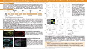
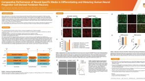 科学海报Comparative Performance of Neural-Specific Media in Differentiating and Maturing Human Neural Progenitor Cell-Derived Forebrain Neurons
Conference:
Society for Neuroscience (SfN)
-
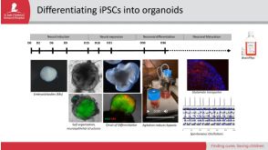
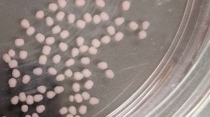 实验方案How to Dissociate 3D Neural Organoids into a Single-Cell Suspension
研究方向:
干细胞生物学,疾病建模,神经科学,类器官,传染病
-
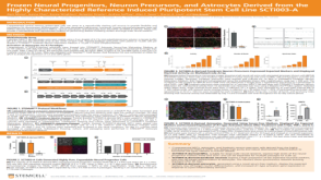
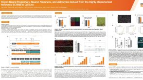 科学海报Frozen Neural Progenitors, Neuron Precursors, and Astrocytes Derived from the Highly Characterized Reference iPSC Line, SCTi003-A
Conference:
Society for Neuroscience (SfN)
-
产品号#:
05835
05839
05854
05855
34811
34815
34821
34825
34850
34860
85850
85857
产品名:
STEMdiff™ 神经诱导培养基
STEMdiff™ 神经诱导培养基
mFreSR™
mFreSR™
AggreWell™ 800 24孔板,1个
AggreWell™ 800 24孔板,5个
AggreWell™ 800 6孔板,1个
AggreWell™ 800 6孔板,5个
AggreWell™ 800 24孔板启动套装
AggreWell™ 800 6孔板启动套装
mTeSR™1
mTeSR™1

 沪公网安备31010102008431号