
 EasySep™小鼠TIL(CD45)正选试剂盒


技术资料
-
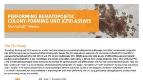
 挂图Identification of Colonies Derived from Mouse Hematopoietic Progenitors Representative colony images and tips for identifying progenitor subtypes in CFU assays
 -

-

-

-

-


 沪公网安备31010102008431号