
 EasySep™小鼠TIL(CD45)正选试剂盒


技术资料
-
-
-
-
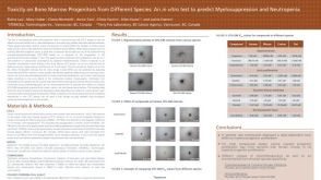
 科学海报Detection of Human Bipotential Erythroid-Megakaryocytic Progenitors in Serum-Free Collagen Gels
Conference:
ISEH 2003
-
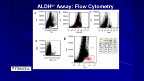
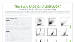 技术公告The Basic FACS on ALDEFLUOR™: The Quick Guide to Flow Cytometry
细胞类型:
乳腺细胞,前列腺细胞,癌细胞及细胞系,脑肿瘤干细胞,造血干/祖细胞
-
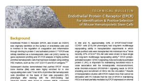
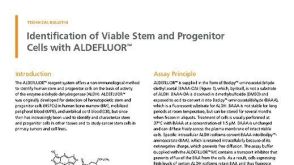 技术公告Identification of Viable Stem and Progenitor Cells with ALDEFLUOR™
细胞类型:
乳腺细胞,前列腺细胞,癌细胞及细胞系,脑肿瘤干细胞,造血干/祖细胞
-
-
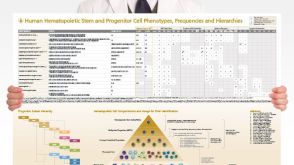 挂图Human Hematopoietic Stem and Progenitor Cell Phenotyping Overview of subset surface markers, frequencies and assays for analysis
 -


 沪公网安备31010102008431号