Arbab AS et al. (SEP 2008)
FASEB journal : official publication of the Federation of American Societies for Experimental Biology 22 9 3234--46
Detection of migration of locally implanted AC133+ stem cells by cellular magnetic resonance imaging with histological findings.
This study investigated the factors responsible for migration and homing of magnetically labeled AC133(+) cells at the sites of active angiogenesis in tumor. AC133(+) cells labeled with ferumoxide-protamine sulfate were mixed with either rat glioma or human melanoma cells and implanted in flank of nude mice. An MRI of the tumors including surrounding tissues was performed. Tumor sections were stained for Prussian blue (PB),platelet-derived growth factor (PDGF),hypoxia-inducible factor-1alpha (HIF-1alpha),stromal cell derived factor-1 (SDF-1),matrix metalloproteinase-2 (MMP-2),vascular endothelial growth factor (VEGF),and endothelial markers. Fresh snap-frozen strips from the central and peripheral parts of the tumor were collected for Western blotting. MRIs demonstrated hypointense regions at the periphery of the tumors where the PB(+)/AC133(+) cells were positive for endothelial cells markers. At the sites of PB(+)/AC133(+) cells,both HIF-1alpha and SDF-1 were strongly positive and PDGF and MMP-2 showed generalized expression in the tumor and surrounding tissues. There was no significant association of PB(+)/AC133(+) cell localization and VEGF expression in tumor cells. Western blot demonstrated strong expression of the SDF-1,MMP-2,and PDGF at the peripheral parts of the tumors. HIF-1alpha was expressed at both the periphery and central parts of the tumor. This work demonstrates that magnetically labeled cells can be used as probes for MRI and histological identification of administered cells.
View Publication

 EasySep™小鼠TIL(CD45)正选试剂盒


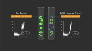
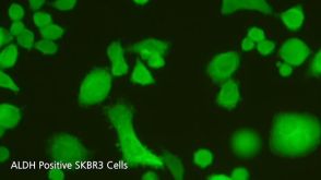
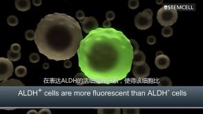
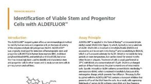 技术公告Identification of Viable Stem and Progenitor Cells with ALDEFLUOR™
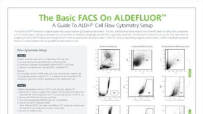 技术公告The Basic FACS on ALDEFLUOR™: The Quick Guide to Flow Cytometry


 沪公网安备31010102008431号