
 EasySep™小鼠TIL(CD45)正选试剂盒


技术资料
-
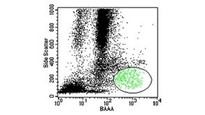
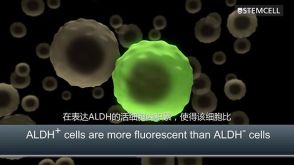
 技术公告Identification of Viable Stem and Progenitor Cells with ALDEFLUOR™
细胞类型:
乳腺细胞,前列腺细胞,癌细胞及细胞系,脑肿瘤干细胞,造血干祖细胞
发布日期: 05/17/2023 -
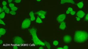
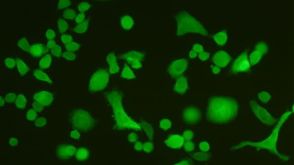
 技术公告The Basic FACS on ALDEFLUOR™: The Quick Guide to Flow Cytometry
细胞类型:
乳腺细胞,前列腺细胞,癌细胞及细胞系,脑肿瘤干细胞,造血干祖细胞
发布日期: 01/23/2019 -

-


 沪公网安备31010102008431号