D. M. Previte et al. (apr 2019)
Cell reports 27 1 129--141.e4
Lymphocyte Activation Gene-3 Maintains Mitochondrial and Metabolic Quiescence in Naive CD4+ T Cells.
Lymphocyte activation gene-3 (LAG-3) is an inhibitory receptor expressed by CD4+ T cells and tempers their homeostatic expansion. Because CD4+ T cell proliferation is tightly coupled to bioenergetics,we investigate the role of LAG-3 in modulating naive CD4+ T cell metabolism. LAG-3 deficiency enhances the metabolic profile of naive CD4+ T cells by elevating levels of mitochondrial biogenesis. In vivo,LAG-3 blockade partially restores expansion and the metabolic phenotype of wild-type CD4+ T cells to levels of Lag3-/- CD4+ T cells,solidifying that LAG-3 controls these processes. Lag3-/- CD4+ T cells also demonstrate greater signal transducer and activator of transcription 5 (STAT5) activation,enabling resistance to interleukin-7 (IL-7) deprivation. These results implicate this pathway as a target of LAG-3-mediated inhibition. Additionally,enhancement of STAT5 activation,as a result of LAG-3 deficiency,contributes to greater activation potential in these cells. These results identify an additional mode of regulation elicited by LAG-3 in controlling CD4+ T cell responses.
View Publication
产品号#:
19852
19852RF
产品名:
EasySep™小鼠CD4+ T细胞分选试剂盒
RoboSep™ 小鼠CD4+ T细胞分选试剂盒
Papait A et al. (NOV 2016)
Journal of tissue engineering and regenerative medicine
Allogeneic platelet-rich plasma affects monocyte differentiation to dendritic cells causing an anti-inflammatory microenvironment putatively fostering the wound healing.
Autologous platelet rich plasma (PRP) is clinically used to induce repair of different tissues through the release of bioactive molecules. In some patients,the production of an efficient autologous PRP is unfeasible due to their compromised health. We developed an allogeneic PRP mismatched for AB0 and Rh antigens. To broadcast its clinical applications avoiding side effects the outcome of allogeneic PRP on immune response should be defined. Thus,we investigated whether PRP affected the differentiation of peripheral blood monocytes to dendritic cells upon stimulation with granulocyte monocyte colony stimulating factor and interleukin-4. Indeed,these cells are the main players of immune response and tissue repair. PRP inhibited the differentiation of monocytes to CD1a(+) dendritic cells and favored the expansion of phagocytic CD163(+) CD206(+) fibrocyte-like cells. These cells produced inteleukin-10 and prostaglandin-E2,but not interferon-γ,upon stimulation with lipopolysaccharides. Moreover,they promoted the expansion of regulatory CD4(+) CD25(+) FoxP3(+) T cells upon allostimulation or antigen specific priming. Finally,the conditioned medium harvested from monocytes differentiated with PRP triggered a strong chemotactic effect on mesenchymal cells in both scratch and transwell migration assays. These results strongly suggest that allogeneic PRP can foster the differentiation of monocytes to a regulatory anti-inflammatory population possibly favoring wound healing.
View Publication
产品号#:
15022
15062
19155
19155RF
15028
15068
产品名:
RosetteSep™人CD4+ T细胞富集抗体混合物
RosetteSep™人CD4+ T细胞富集抗体混合物
RosetteSep™人单核细胞富集抗体混合物
RosetteSep™人单核细胞富集抗体混合物
Li R et al. (NOV 2016)
Cancer research
Macrophage-secreted TNFα and TGFβ1 Influence Migration Speed and Persistence of Cancer Cells in 3D Tissue Culture via Independent Pathways.
The ability of a cancer cell to migrate through the dense extracellular matrix (ECM) within and surrounding the solid tumor is a critical determinant of metastasis. Macrophages enhance invasion and metastasis in the tumor microenvironment but the basis for their effects are not fully understood. Using a microfluidic 3D cell migration assay,we found that the presence of macrophages enhanced the speed and persistence of cancer cell migration through a 3D extracellular matrix in a matrix metalloproteinases (MMP)-dependent fashion. Mechanistic investigations revealed that macrophage-released TNFα and TGFβ1 mediated the observed behaviors by two distinct pathways. These factors synergistically enhanced migration persistence through a synergistic induction of NF-κB-dependent MMP1 expression in cancer cells. In contrast,macrophage-released TGFβ1 enhanced migration speed primarily by inducing MT1-MMP expression. Taken together,our results reveal new insights into how macrophages enhance cancer cell metastasis,and they identify TNFα and TGFβ1 dual blockade as an anti-metastatic strategy in solid tumors.
View Publication
产品号#:
19059
19059RF
产品名:
EasySep™人单核细胞富集试剂盒
RoboSep™ 人单核细胞富集试剂盒含滤芯吸头
Loo CP et al. (NOV 2016)
Journal of immunology (Baltimore,Md. : 1950)
Blocking Virus Replication during Acute Murine Cytomegalovirus Infection Paradoxically Prolongs Antigen Presentation and Increases the CD8+ T Cell Response by Preventing Type I IFN-Dependent Depletion of Dendritic Cells.
Increasing amounts of pathogen replication usually lead to a proportionate increase in size and effector differentiation of the CD8(+) T cell response,which is attributed to increased Ag and inflammation. Using a murine CMV that is highly sensitive to the antiviral drug famciclovir to modulate virus replication,we found that increased virus replication drove increased effector CD8(+) T cell differentiation,as expected. Paradoxically,however,increased virus replication dramatically decreased the size of the CD8(+) T cell response to two immunodominant epitopes. The decreased response was due to type I IFN-dependent depletion of conventional dendritic cells and could be reproduced by specific depletion of dendritic cells from day 2 postinfection or by sterile induction of type I IFN. Increased virus replication and type I IFN specifically inhibited the response to two immunodominant epitopes that are known to be dependent on Ag cross-presented by DCs,but they did not inhibit the response to inflationary" epitopes whose responses can be sustained by infected nonhematopoietic cells. Our results show that type I IFN can suppress CD8(+) T cell responses to cross-presented Ag by depleting cross-presenting conventional dendritic cells."
View Publication
产品号#:
19853
19853RF
产品名:
EasySep™小鼠CD8+ T细胞分选试剂盒
RoboSep™ 小鼠CD8+ T细胞分选试剂盒
Figueroa G et al. (OCT 2016)
Journal of visualized experiments : JoVE 116
Characterization of Human Monocyte-derived Dendritic Cells by Imaging Flow Cytometry: A Comparison between Two Monocyte Isolation Protocols.
Dendritic cells (DCs) are antigen presenting cells of the immune system that play a crucial role in lymphocyte responses,host defense mechanisms,and pathogenesis of inflammation. Isolation and study of DCs have been important in biological research because of their distinctive features. Although they are essential key mediators of the immune system,DCs are very rare in blood,accounting for approximately 0.1 - 1% of total blood mononuclear cells. Therefore,alternatives for isolation methods rely on the differentiation of DCs from monocytes isolated from peripheral blood mononuclear cells (PBMCs). The utilization of proper isolation techniques that combine simplicity,affordability,high purity,and high yield of cells is imperative to consider. In the current study,two distinct methods for the generation of DCs will be compared. Monocytes were selected by adherence or negatively enriched using magnetic separation procedure followed by differentiation into DCs with IL-4 and GM-CSF. Monocyte and MDDC viability,proliferation,and phenotype were assessed using viability dyes,MTT assay,and CD11c/ CD14 surface marker analysis by imaging flow cytometry. Although the magnetic separation method yielded a significant higher percentage of monocytes with higher proliferative capacity when compared to the adhesion method,the findings have demonstrated the ability of both techniques to simultaneously generate monocytes that are capable of proliferating and differentiating into viable CD11c+ MDDCs after seven days in culture. Both methods yielded textgreater 70% CD11c+ MDDCs. Therefore,our results provide insights that contribute to the development of reliable methods for isolation and characterization of human DCs.
View Publication

 EasySep™小鼠TIL(CD45)正选试剂盒


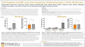 科学海报Centrifugation and RBC Lysis-Free Preparation of Blood Samples in Under 30 Minutes
 实验方案How to Process Leukocyte Reduction System (LRS) Cones/Chambers for Downstream Cell Isolation

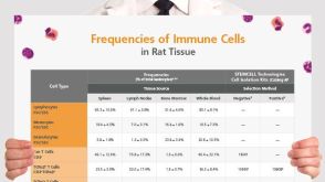 挂图Frequencies of Immune Cells in Rat Tissue Lists the estimated frequencies of more than 15 immune cell types in Sprague Dawley rats

 沪公网安备31010102008431号