Khazen R et al. (MAR 2016)
Nature Communications 7 10823
Melanoma cell lysosome secretory burst neutralizes the CTL-mediated cytotoxicity at the lytic synapse.
Human melanoma cells express various tumour antigens that are recognized by CD8(+) cytotoxic T lymphocytes (CTLs) and elicit tumour-specific responses in vivo. However,natural and therapeutically enhanced CTL responses in melanoma patients are of limited efficacy. The mechanisms underlying CTL effector phase failure when facing melanomas are still largely elusive. Here we show that,on conjugation with CTL,human melanoma cells undergo an active late endosome/lysosome trafficking,which is intensified at the lytic synapse and is paralleled by cathepsin-mediated perforin degradation and deficient granzyme B penetration. Abortion of SNAP-23-dependent lysosomal trafficking,pH perturbation or impairment of lysosomal proteolytic activity restores susceptibility to CTL attack. Inside the arsenal of melanoma cell strategies to escape immune surveillance,we identify a self-defence mechanism based on exacerbated lysosome secretion and perforin degradation at the lytic synapse. Interfering with this synaptic self-defence mechanism might be useful in potentiating CTL-mediated therapies in melanoma patients.
View Publication
Bosma M et al. (APR 2016)
Nature Communications 7 11314
FNDC4 acts as an anti-inflammatory factor on macrophages and improves colitis in mice.
FNDC4 is a secreted factor sharing high homology with the exercise-associated myokine irisin (FNDC5). Here we report that Fndc4 is robustly upregulated in several mouse models of inflammation as well as in human inflammatory conditions. Specifically,FNDC4 levels are increased locally at inflamed sites of the intestine of inflammatory bowel disease patients. Interestingly,administration of recombinant FNDC4 in the mouse model of induced colitis markedly reduces disease severity compared with mice injected with a control protein. Conversely,mice lacking Fndc4 develop more severe colitis. Analysis of binding of FNDC4 to different immune cell types reveals strong and specific binding to macrophages and monocytes. FNDC4 treatment of bone marrow-derived macrophages in vitro results in reduced phagocytosis,increased cell survival and reduced proinflammatory chemokine expression. Hence,treatment with FNDC4 results in a state of dampened macrophage activity,while enhancing their survival. Thus,we have characterized FNDC4 as a factor with direct therapeutic potential in inflammatory bowel disease and possibly other inflammatory diseases.
View Publication
产品号#:
19359
19359RF
100-0697
产品名:
EasySep™人单核细胞分选试剂盒
RoboSep™ 人单核细胞分选试剂盒
EasySep™人单核细胞分选试剂盒
de Valle E et al. (APR 2016)
The Journal of Experimental Medicine 213 4 621--41
NFκB1 is essential to prevent the development of multiorgan autoimmunity by limiting IL-6 production in follicular B cells.
We examined the role of NFκB1 in the homeostasis and function of peripheral follicular (Fo) B cells. Aging mice lacking NFκB1 (Nfκb1(-/-)) develop lymphoproliferative and multiorgan autoimmune disease attributed in large part to the deregulated activity ofNfκb1(-/-)Fo B cells that produce excessive levels of the proinflammatory cytokine interleukin 6 (IL-6). Despite enhanced germinal center (GC) B cell differentiation,the formation of GC structures was severely disrupted in theNfκb1(-/-)mice. Bone marrow chimeric mice revealed that the Fo B cell-intrinsic loss of NFκB1 led to the spontaneous generation of GC B cells. This was primarily the result of an increase in IL-6 levels,which promotes the differentiation of Fo helper CD4(+)T cells and acts in an autocrine manner to reduce antigen receptor and toll-like receptor activation thresholds in a population of proliferating IgM(+)Nfκb1(-/-)Fo B cells. We demonstrate that p50-NFκB1 repressesIl-6transcription in Fo B cells,with the loss of NFκB1 also resulting in the uncontrolled RELA-driven transcription ofIl-6.Collectively,our findings identify a previously unrecognized role for NFκB1 in preventing multiorgan autoimmunity through its negative regulation ofIl-6gene expression in Fo B cells.
View Publication
产品号#:
19854
19854RF
产品名:
EasySep™小鼠B细胞分选试剂盒
RoboSep™ 小鼠B细胞分选试剂盒
Vasu S et al. (MAR 2016)
Blood
Decitabine enhances Fc engineered anti-CD33 mAb mediated natural killer antibody dependent cellular cytotoxicity against AML blasts.
Acute myeloid leukemia (AML) is the most common type of acute leukemia affecting older individuals at a median age of 67 years. Resistance to intensive induction chemotherapy is the major cause of death in elderly AML; hence novel treatment strategies are warranted. CD33-directed antibody-drug conjugates (Gemtuzumab ozogamicin) have been shown to improve overall survival,validating CD33 as a target for antibody-based therapy of AML. Here we report the in vitro efficacy of BI 836858,a fully human,Fc-engineered,anti-CD33 antibody using AML cell lines and primary AML blasts as targets. BI 836858-opsonized AML cells significantly induced both autologous and allogeneic natural killer (NK)-cell degranulation and NK cell-mediated antibody-dependent cellular cytotoxicity (ADCC). In vitro treatment of AML blasts with decitabine (DAC) or 5-azacytidine,two hypomethylating agents that show efficacy in older patients,did not compromise BI 836858-induced NK cell-mediated ADCC. Evaluation of BI 836858-mediated ADCC in serial marrow AML aspirates in patients who received a ten-day course of DAC (pre-DAC,days 4,11 and 28 post-DAC) revealed significantly higher ADCC in samples at day 28 post-DAC when compared to pre-DAC treatment. Analysis of ligands (L) to activating receptors (NKG2D showed significantly increased NKG2DL expression in day 28 post-DAC samples compared to pre-DAC samples; when NKG2DL receptor was blocked using antibodies,BI 836858-mediated ADCC was significantly decreased,suggesting that DAC enhances AML blast susceptibility to BI 836858 by upregulating NKG2DL. These data provide a rationale for combination therapy of Fc-engineered antibodies such as BI 836858 with azanucleosides in elderly patients with AML.
View Publication
产品号#:
15025
15065
产品名:
RosetteSep™人NK细胞富集抗体混合物
RosetteSep™人NK细胞富集抗体混合物
Y. Kuwano et al. (MAY 2016)
Journal of Immunology 196 9 3828--33
G$\alpha$i2 and G$\alpha$i3 Differentially Regulate Arrest from Flow and Chemotaxis in Mouse Neutrophils.
Leukocyte recruitment to inflammation sites progresses in a multistep cascade. Chemokines regulate multiple steps of the cascade,including arrest,transmigration,and chemotaxis. The most important chemokine receptor in mouse neutrophils is CXCR2,which couples through G$\alpha$i2- and G$\alpha$i3-containing heterotrimeric G proteins. Neutrophils arrest in response to CXCR2 stimulation. This is defective in G$\alpha$i2-deficient neutrophils. In this study,we show that G$\alpha$i3-deficient neutrophils showed reduced transmigration but normal arrest in mice. We also tested G$\alpha$i2- or G$\alpha$i3-deficient neutrophils in a CXCL1 gradient generated by a microfluidic device. G$\alpha$i3-,but not G$\alpha$i2-,deficient neutrophils showed significantly reduced migration and directionality. This was confirmed in a model of sterile inflammation in vivo. G$\alpha$i2-,but not G$\alpha$i3-,deficient neutrophils showed decreased Ca(2+) flux in response to CXCR2 stimulation. Conversely,G$\alpha$i3-,but not G$\alpha$i2-,deficient neutrophils exhibited reduced AKT phosphorylation upon CXCR2 stimulation. We conclude that G$\alpha$i2 controls arrest and G$\alpha$i3 controls transmigration and chemotaxis in response to chemokine stimulation of neutrophils.
View Publication
产品号#:
19762
19762RF
20155
产品名:
EasySep™小鼠中性粒细胞富集试剂盒
RoboSep™ 小鼠中性粒细胞富集试剂盒含滤芯吸头
RoboSep™分选管套装(9个塑料管)
Serr I et al. (MAR 2016)
Nature Communications 7 10991
Type 1 diabetes vaccine candidates promote human Foxp3(+)Treg induction in humanized mice.
Immune tolerance is executed partly by Foxp3(+)regulatory T (Treg) cells,which suppress autoreactive T cells. In autoimmune type 1 diabetes (T1D) impaired tolerance promotes destruction of insulin-producing β-cells. The development of autoantigen-specific vaccination strategies for Foxp3(+)Treg-induction and prevention of islet autoimmunity in patients is still in its infancy. Here,using human haematopoietic stem cell-engrafted NSG-HLA-DQ8 transgenic mice,we provide direct evidence for human autoantigen-specific Foxp3(+)Treg-induction in vivo. We identify HLA-DQ8-restricted insulin-specific CD4(+)T cells and demonstrate efficient human insulin-specific Foxp3(+)Treg-induction upon subimmunogenic vaccination with strong agonistic insulin mimetopes in vivo. Induced human Tregs are stable,show increased expression of Treg signature genes such as Foxp3,CTLA4,IL-2Rα and TIGIT and can efficiently suppress effector T cells. Such Foxp3(+)Treg-induction does not trigger any effector T cells. These T1D vaccine candidates could therefore represent an expedient improvement in the challenge to induce human Foxp3(+)Tregs and to develop novel precision medicines for prevention of islet autoimmunity in children at risk of T1D.
View Publication
产品号#:
17952
17952RF
100-0696
产品名:
EasySep™人CD4+ T细胞分选试剂盒
RoboSep™ 人CD4+ T细胞分选试剂盒
EasySep™人CD4+ T细胞分离试剂盒
Flach A-C et al. (MAR 2016)
Proceedings of the National Academy of Sciences of the United States of America 113 12 3323--8
Autoantibody-boosted T-cell reactivation in the target organ triggers manifestation of autoimmune CNS disease.
Multiple sclerosis (MS) is caused by T cells that are reactive for brain antigens. In experimental autoimmune encephalomyelitis,the animal model for MS,myelin-reactive T cells initiate the autoimmune process when entering the nervous tissue and become reactivated upon local encounter of their cognate CNS antigen. Thereby,the strength of the T-cellular reactivation process within the CNS tissue is crucial for the manifestation and the severity of the clinical disease. Recently,B cells were found to participate in the pathogenesis of CNS autoimmunity,with several diverse underlying mechanisms being under discussion. We here report that B cells play an important role in promoting the initiation process of CNS autoimmunity. Myelin-specific antibodies produced by autoreactive B cells after activation in the periphery diffused into the CNS together with the first invading pathogenic T cells. The antibodies accumulated in resident antigen-presenting phagocytes and significantly enhanced the activation of the incoming effector T cells. The ensuing strong blood-brain barrier disruption and immune cell recruitment resulted in rapid manifestation of clinical disease. Therefore,myelin oligodendrocyte glycoprotein (MOG)-specific autoantibodies can initiate disease bouts by cooperating with the autoreactive T cells in helping them to recognize their autoantigen and become efficiently reactivated within the immune-deprived nervous tissue.
View Publication
产品号#:
19851
19851RF
19854
19854RF
产品名:
EasySep™小鼠T细胞分选试剂盒
RoboSep™ 小鼠T细胞分选试剂盒
EasySep™小鼠B细胞分选试剂盒
RoboSep™ 小鼠B细胞分选试剂盒
Gracias DT et al. (FEB 2016)
Journal of Immunology 196 3 1186--98
Phosphatidylinositol 3-Kinase p110δ Isoform Regulates CD8+ T Cell Responses during Acute Viral and Intracellular Bacterial Infections.
The p110δ isoform of PI3K is known to play an important role in immunity,yet its contribution to CTL responses has not been fully elucidated. Using murine p110δ-deficient CD8(+) T cells,we demonstrated a critical role for the p110δ subunit in the generation of optimal primary and memory CD8(+) T cell responses. This was demonstrated in both acute viral and intracellular bacterial infections in mice. We show that p110δ signaling is required for CD8(+) T cell activation,proliferation and effector cytokine production. We provide evidence that the effects of p110δ signaling are mediated via Akt activation and through the regulation of TCR-activated oxidative phosphorylation and aerobic glycolysis. In light of recent clinical trials that employ drugs targeting p110δ in certain cancers and other diseases,our study suggests caution in using these drugs in patients,as they could potentially increase susceptibility to infectious diseases. These studies therefore reveal a novel and direct role for p110δ signaling in in vivo CD8(+) T cell immunity to microbial pathogens.
View Publication

 EasySep™小鼠TIL(CD45)正选试剂盒


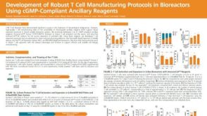 科学海报Development of Robust T Cell Manufacturing Protocols in Bioreactors Using cGMP-Compliant Ancillary Reagents

 沪公网安备31010102008431号