Locally produced C5a binds to T cell-expressed C5aR to enhance effector T-cell expansion by limiting antigen-induced apoptosis.
Our recent studies have shown that immune cell-produced complement provides costimulatory and survival signals to naive CD4(+) T cells. Whether these signals are similarly required during effector cell expansion and what molecular pathways link locally produced complement to T-cell survival were not clarified. To address this,we stimulated monoclonal and polyclonal T cells in vitro and in vivo with antigen-presenting cells (APCs) deficient in the complement regulatory protein,decay accelerating factor (DAF),and/or the complement component C3. We found that T-cell expansion induced by DAF-deficient APCs was augmented with diminished T-cell apoptosis,whereas T-cell expansion induced by C3(-/-) APCs was reduced because of enhanced T-cell apoptosis. These effects were traced to locally produced C5a,which through binding to T cell-expressed C5aR,enhanced expression of Bcl-2 and prevented Fas up-regulation. The results show that C5aR signal transduction in T cells is important to allow optimal T-cell expansion,as well as to maintain naive cell viability,and does so by suppressing programmed cell death.
View Publication
产品号#:
19751
19751RF
产品名:
Maldonado RA et al. (APR 2009)
The Journal of experimental medicine 206 4 877--92
Control of T helper cell differentiation through cytokine receptor inclusion in the immunological synapse.
The antigen recognition interface formed by T helper precursors (Thps) and antigen-presenting cells (APCs),called the immunological synapse (IS),includes receptors and signaling molecules necessary for Thp activation and differentiation. We have recently shown that recruitment of the interferon-gamma receptor (IFNGR) into the IS correlates with the capacity of Thps to differentiate into Th1 effector cells,an event regulated by signaling through the functionally opposing receptor to interleukin-4 (IL4R). Here,we show that,similar to IFN-gamma ligation,TCR stimuli induce the translocation of signal transducer and activator of transcription 1 (STAT1) to IFNGR1-rich regions of the membrane. Unexpectedly,STAT1 is preferentially expressed,is constitutively serine (727) phosphorylated in Thp,and is recruited to the IS and the nucleus upon TCR signaling. IL4R engagement controls this process by interfering with both STAT1 recruitment and nuclear translocation. We also show that in cells with deficient Th1 or constitutive Th2 differentiation,the IL4R is recruited to the IS. This observation suggest that the IL4R is retained outside the IS,similar to the exclusion of IFNGR from the IS during IL4R signaling. This study provides new mechanistic cues for the regulation of lineage commitment by mutual immobilization of functionally antagonistic membrane receptors.
View Publication
产品号#:
21000
20119
20155
19752
19752RF
产品名:
RoboSep™- S
RoboSep™ 吸头组件抛光剂
RoboSep™分选管套装(9个塑料管)
Wang X et al. (MAR 2009)
Journal of immunology (Baltimore,Md. : 1950) 182 6 3597--608
MEKK3 is essential for lymphopenia-induced T cell proliferation and survival.
T cell homeostasis is crucial for maintaining an efficient and balanced T cell immunity. The interaction between TCR and self peptide (sp) MHC ligands is known to be the key driving force in this process,and it is believed to be functionally and mechanistically different from that initiated by the antigenic TCR stimulation. Yet,very little is known about the downstream signaling events triggered by this TCR-spMHC interaction and how they differ from those triggered by antigenic TCR stimulation. In this study,we show that T cell conditional ablation of MEKK3,a Ser/Thr kinase in the MAPK cascade,causes a significant reduction in peripheral T cell numbers in the conditional knockout mice,but does not perturb thymic T cell development and maturation. Using an adoptive mixed transfer method,we show that MEKK3-deficient T cells are severely impaired in lymphopenia-induced cell proliferation and survival. Interestingly,the Ag-induced T cell proliferation proceeds normally in the absence of MEKK3. Finally,we found that the activity of ERK1/2,but not p38 MAPK,was attenuated during the lymphopenia-driven response in MEKK3-deficient T cells. Together,these data suggest that MEKK3 may play a crucial selective role for spMHC-mediated T cell homeostasis.
View Publication
产品号#:
18751
18751RF
产品名:
Popovic R et al. (APR 2009)
Blood 113 14 3314--22
Regulation of mir-196b by MLL and its overexpression by MLL fusions contributes to immortalization.
Chromosomal translocations involving the Mixed Lineage Leukemia (MLL) gene produce chimeric proteins that cause abnormal expression of a subset of HOX genes and leukemia development. Here,we show that MLL normally regulates expression of mir-196b,a hematopoietic microRNA located within the HoxA cluster,in a pattern similar to that of the surrounding 5' Hox genes,Hoxa9 and Hoxa10,during embryonic stem (ES) cell differentiation. Within the hematopoietic lineage,mir-196b is most abundant in short-term hematopoietic stem cells and is down-regulated in more differentiated hematopoietic cells. Leukemogenic MLL fusion proteins cause overexpression of mir-196b,while treatment of MLL-AF9 transformed bone marrow cells with mir-196-specific antagomir abrogates their replating potential in methylcellulose. This demonstrates that mir-196b function is necessary for MLL fusion-mediated immortalization. Furthermore,overexpression of mir-196b was found specifically in patients with MLL associated leukemias as determined from analysis of 55 primary leukemia samples. Overexpression of mir-196b in bone marrow progenitor cells leads to increased proliferative capacity and survival,as well as a partial block in differentiation. Our results suggest a mechanism whereby increased expression of mir-196b by MLL fusion proteins significantly contributes to leukemia development.
View Publication
产品号#:
03534
19756
19756RF
产品名:
MethoCult™ GF M3534
Imbeault M et al. (JAN 2009)
Retrovirology 6 5
Microarray study reveals that HIV-1 induces rapid type-I interferon-dependent p53 mRNA up-regulation in human primary CD4+ T cells.
BACKGROUND: Infection with HIV-1 has been shown to alter expression of a large array of host cell genes. However,previous studies aimed at investigating the putative HIV-1-induced modulation of host gene expression have been mostly performed in established human cell lines. To better approximate natural conditions,we monitored gene expression changes in a cell population highly enriched in human primary CD4+ T lymphocytes exposed to HIV-1 using commercial oligonucleotide microarrays from Affymetrix. RESULTS: We report here that HIV-1 influences expression of genes related to many important biological processes such as DNA repair,cellular cycle,RNA metabolism and apoptosis. Notably,expression of the p53 tumor suppressor and genes involved in p53 homeostasis such as GADD34 were up-regulated by HIV-1 at the mRNA level. This observation is distinct from the previously reported p53 phosphorylation and stabilization at the protein level,which precedes HIV-1-induced apoptosis. We present evidence that the HIV-1-mediated increase in p53 gene expression is associated with virus-mediated induction of type-I interferon (i.e. IFN-alpha and IFN-beta). CONCLUSION: These observations have important implications for our understanding of HIV-1 pathogenesis,particularly in respect to the virus-induced depletion of CD4+ T cells.
View Publication
Engineering a stable and selective peptide blocker of the Kv1.3 channel in T lymphocytes.
Kv1.3 potassium channels maintain the membrane potential of effector memory (T(EM)) T cells that are important mediators of multiple sclerosis,type 1 diabetes mellitus,and rheumatoid arthritis. The polypeptide ShK-170 (ShK-L5),containing an N-terminal phosphotyrosine extension of the Stichodactyla helianthus ShK toxin,is a potent and selective blocker of these channels. However,a stability study of ShK-170 showed minor pH-related hydrolysis and oxidation byproducts that were exacerbated by increasing temperatures. We therefore engineered a series of analogs to minimize the formation of these byproducts. The analog with the greatest stability,ShK-192,contains a nonhydrolyzable phosphotyrosine surrogate,a methionine isostere,and a C-terminal amide. ShK-192 shows the same overall fold as ShK,and there is no evidence of any interaction between the N-terminal adduct and the rest of the peptide. The docking configuration of ShK-192 in Kv1.3 shows the N-terminal para-phosphonophenylalanine group lying at the junction of two channel monomers to form a salt bridge with Lys(411) of the channel. ShK-192 blocks Kv1.3 with an IC(50) of 140 pM and exhibits greater than 100-fold selectivity over closely related channels. After a single subcutaneous injection of 100 microg/kg,approximately 100 to 200 pM concentrations of active peptide is detectable in the blood of Lewis rats 24,48,and 72 h after the injection. ShK-192 effectively inhibits the proliferation of T(EM) cells and suppresses delayed type hypersensitivity when administered at 10 or 100 microg/kg by subcutaneous injection once daily. ShK-192 has potential as a therapeutic for autoimmune diseases mediated by T(EM) cells.
View Publication
产品号#:
19051
19051RF
产品名:
EasySep™人T细胞富集试剂盒
RoboSep™ 人T细胞富集试剂盒含滤芯吸头
Peterson ME and Long EO (OCT 2008)
Immunity 29 4 578--88
Inhibitory receptor signaling via tyrosine phosphorylation of the adaptor Crk.
Many cellular responses,such as autoimmunity and cytotoxicity,are controlled by receptors with cytoplasmic immunoreceptor tyrosine-based inhibition motifs (ITIMs). Here,we showed that binding of inhibitory natural killer (NK) cell receptors to human leukocyte antigen (HLA) class I on target cells induced tyrosine phosphorylation of the adaptor Crk,concomitant with dephosphorylation of the guanine exchange factor Vav1. Furthermore,Crk dissociated from the guanine exchange factor C3G and bound to the tyrosine kinase c-Abl during inhibition. Membrane targeting of a tyrosine-mutated form of Crk could overcome inhibition of NK cell cytotoxicity,providing functional evidence that Crk phosphorylation contributes to inhibition. The specific phosphorylation of Crk and its dissociation from a signaling complex,observed here with two types of inhibitory receptors,expands the signaling potential of the large ITIM-receptor family and reveals an unsuspected component of the inhibitory mechanism.
View Publication
产品号#:
05150
产品名:
MyeloCult™ H5100
Schü et al. (MAY 2008)
Blood 111 9 4532--41
The MADS transcription factor Mef2c is a pivotal modulator of myeloid cell fate.
Mef2c is a MADS (MCM1-agamous-deficient serum response factor) transcription factor best known for its role in muscle and cardiovascular development. A causal role of up-regulated MEF2C expression in myelomonocytic acute myeloid leukemia (AML) has recently been demonstrated. Due to the pronounced monocytic component observed in Mef2c-induced AML,this study was designed to assess the importance of Mef2c in normal myeloid differentiation. Analysis of bone marrow (BM) cells manipulated to constitutively express Mef2c demonstrated increased monopoiesis at the expense of granulopoiesis,whereas BM isolated from Mef2c(Delta/-) mice showed reduced levels of monocytic differentiation in response to cytokines. Mechanistic studies showed that loss of Mef2c expression correlated with reduced levels of transcripts encoding c-Jun,but not PU.1,C/EBPalpha,or JunB transcription factors. Inhibiting Jun expression by short-interfering RNA impaired Mef2c-mediated inhibition of granulocyte development. Moreover,retroviral expression of c-Jun in BM cells promoted monocytic differentiation. The ability of Mef2c to modulate cell-fate decisions between monocyte and granulocyte differentiation,coupled with its functional sensitivity to extracellular stimuli,demonstrate an important role in immunity--and,consistent with findings of other myeloid transcription factors,a target of oncogenic lesions in AML.
View Publication
产品号#:
03434
03444
09600
09650
18556
18556RF
产品名:
MethoCult™ GF M3434
MethoCult™ GF M3434
StemSpan™ SFEM
StemSpan™ SFEM
Chua KY et al. (JAN 2008)
Methods in molecular biology (Clifton,N.J.) 423 509--20
Production of monoclonal antibody by DNA immunization with electroporation.
DNA immunization with in vivo electroporation is an efficient alternative protocol for the production of monoclonal antibodies (mAb). Generation of mAb by DNA immunization is a novel approach to circumvent the following technical hurdles associated with problematic antigens: low abundance and protein instability and use of recombinant proteins that lack posttranslational modifications. This chapter describes the use of a DNA-based immunization protocol for the production of mAb against a house dust mite allergen,designated as Blo t 11,which is a paramyosin homologue found in Blomia tropicalis mites. The Blo t 11 cDNA fused at the N terminus to the sequence of a signal peptide was cloned into the pCI mammalian expression vector. The DNA construct was injected intramuscularly with in vivo electroporation into mice,and the specific antibody production in mice was analyzed by enzyme-linked immunosorbent assay (ELISA). Hybridomas were generated by fusing mouse splenocytes with myeloma cells using the ClonaCell-HY Hybridoma Cloning Kit. Six hybridoma clones secreting Blo t 11 mAb were successfully generated,and these mAb are useful reagents for immunoaffinity purification and immunoassays.
View Publication

 EasySep™小鼠TIL(CD45)正选试剂盒


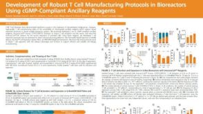 科学海报Development of Robust T Cell Manufacturing Protocols in Bioreactors Using cGMP-Compliant Ancillary Reagents
 沪公网安备31010102008431号