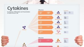
Human Immune Cytokines
Infographic of key cytokines for expansion, differentiation and characterization of major immune cell types
Miyoshi H et al. (JAN 1999)
Science (New York,N.Y.) 283 5402 682--6
Transduction of human CD34+ cells that mediate long-term engraftment of NOD/SCID mice by HIV vectors.
Efficient gene transfer into human hematopoietic stem cells (HSCs) is an important goal in the study of the hematopoietic system as well as for gene therapy of hematopoietic disorders. A lentiviral vector based on the human immunodeficiency virus (HIV) was able to transduce human CD34+ cells capable of stable,long-term reconstitution of nonobese diabetic/severe combined immunodeficient (NOD/SCID) mice. High-efficiency transduction occurred in the absence of cytokine stimulation and resulted in transgene expression in multiple lineages of human hematopoietic cells for up to 22 weeks after transplantation.
View Publication
产品号#:
09500
产品名:
BIT 9500血清替代物
D. Xie et al. (MAY 2017)
Experimental cell research
The effects of activin A on the migration of human breast cancer cells and neutrophils and their migratory interaction.
Activin A belongs to the superfamily of transforming growth factor beta (TGF$\beta$) and is a critical regulatory cytokine in breast cancer and inflammation. However,the role of activin A in migration of breast cancer cells and immune cells was not well characterized. Here,a microfluidic device was used to examine the effect of activin A on the migration of human breast cancer cell line MDA-MB-231 cells and human blood neutrophils as well as their migratory interaction. We found that activin A promoted the basal migration but impaired epidermal growth factor (EGF)-induced migration of breast cancer cells. By contrast,activin A reduced neutrophil chemotaxis and transendothelial migration to N-Formyl-Met-Leu-Phe (fMLP). Finally,activin A promoted neutrophil chemotaxis to the supernatant from breast cancer cell culture. Collectively,our study revealed the different roles of activin A in regulating the migration of breast cancer cells and neutrophils and their migratory interaction. These findings suggested the potential of activin A as a therapeutic target for inflammation and breast cancers.
View Publication
产品号#:
19666
100-0404
产品名:
EasySep™ Direct人中性粒细胞分选试剂盒
RoboSep™ 人中性粒细胞分选试剂盒
Liu Y-S et al. (MAY 2017)
Oncogene
MiR-181b modulates EGFR-dependent VCAM-1 expression and monocyte adhesion in glioblastoma.
Tumor-associated macrophages (TAMs) originate as circulating monocytes,and are recruited to gliomas,where they facilitate tumor growth and migration. Understanding the interaction between TAM and cancer cells may identify therapeutic targets for glioblastoma multiforme (GBM). Vascular cell adhesion molecule-1 (VCAM-1) is a cytokine-induced adhesion molecule expressed on the surface of cancer cells,which is involved in interactions with immune cells. Analysis of the glioma patient database and tissue immunohistochemistry showed that VCAM-1 expression correlated with the clinico-pathological grade of gliomas. Here,we found that VCAM-1 expression correlated positively with monocyte adhesion to GBM,and knockdown of VCAM-1 abolished the enhancement of monocyte adhesion. Importantly,upregulation of VCAM-1 is dependent on epidermal-growth-factor-receptor (EGFR) expression,and inhibition of EGFR effectively reduced VCAM-1 expression and monocyte adhesion activity. Moreover,GBM possessing higher EGFR levels (U251 cells) had higher VCAM-1 levels compared to GBMs with lower levels of EGFR (GL261 cells). Using two- and three-dimensional cultures,we found that monocyte adhesion to GBM occurs via integrin α4β1,which promotes tumor growth and invasion activity. Increased proliferation and tumor necrosis factor-α and IFN-γ levels were also observed in the adherent monocytes. Using a genetic modification approach,we demonstrated that VCAM-1 expression and monocyte adhesion were regulated by the miR-181 family,and lower levels of miR-181b correlated with high-grade glioma patients. Our results also demonstrated that miR-181b/protein phosphatase 2A-modulated SP-1 de-phosphorylation,which mediated the EGFR-dependent VCAM-1 expression and monocyte adhesion to GBM. We also found that the EGFR-dependent VCAM-1 expression is mediated by the p38/STAT3 signaling pathway. Our study suggested that VCAM-1 is a critical modulator of EGFR-dependent interaction of monocytes with GBM,which raises the possibility of developing effective and improved therapies for GBM.Oncogene advance online publication,1 May 2017; doi:10.1038/onc.2017.129.
View Publication
产品号#:
15028
15068
产品名:
RosetteSep™人单核细胞富集抗体混合物
RosetteSep™人单核细胞富集抗体混合物
Fu W et al. (DEC 2016)
Scientific reports 6 38162
Immune Activation Influences SAMHD1 Expression and Vpx-mediated SAMHD1 Degradation during Chronic HIV-1 Infection.
SAMHD1 restricts human immunodeficiency virus type 1 (HIV-1) replication in myeloid cells and CD4(+) T cells,while Vpx can mediate SAMHD1 degradation to promote HIV-1 replication. Although the restriction mechanisms of SAMHD1 have been well-described,SAMHD1 expression and Vpx-mediated SAMHD1 degradation during chronic HIV-1 infection were poorly understood. Flow cytometric analysis was used to directly visualize ex vivo,and after in vitro SIV-Vpx treatment,SAMHD1 expression in CD4(+) T cells and monocytes. Here we report activated CD4(+) T cells without SAMHD1 expression were severely reduced,and SAMHD1 in CD4(+) T cells became susceptible to SIV-Vpx mediated degradation during chronic HIV-1 infection,which was absent from uninfected donors. These alterations were irreversible,even after long-term fully suppressive antiretroviral treatment. Although SAMHD1 expression in CD4(+) T cells and monocytes was not found to correlate with plasma viral load,Vpx-mediated SAMHD1 degradation was associated with indicators of immune activation. In vitro assays further revealed that T-cell activation and an upregulated IFN-I pathway contributed to these altered SAMHD1 properties. These findings provide insight into how immune activation during HIV-1 infection leads to irreparable aberrations in restriction factors and in subsequent viral evasion from host antiviral defenses.
View Publication
产品号#:
17952
17952RF
19359
19359RF
100-0696
100-0697
产品名:
EasySep™人CD4+ T细胞分选试剂盒
RoboSep™ 人CD4+ T细胞分选试剂盒
EasySep™人单核细胞分选试剂盒
RoboSep™ 人单核细胞分选试剂盒
EasySep™人CD4+ T细胞分离试剂盒
EasySep™人单核细胞分选试剂盒
Donnarumma T et al. (NOV 2016)
Cell reports 17 6 1571--1583
Opposing Development of Cytotoxic and Follicular Helper CD4 T Cells Controlled by the TCF-1-Bcl6 Nexus.
CD4(+) T cells develop distinct and often contrasting helper,regulatory,or cytotoxic activities. Typically a property of CD8(+) T cells,granzyme-mediated cytotoxic T cell (CTL) potential is also exerted by CD4(+) T cells. However,the conditions that induce CD4(+) CTLs are not entirely understood. Using single-cell transcriptional profiling,we uncover a unique signature of Granzyme B (GzmB)(+) CD4(+) CTLs,which distinguishes them from other CD4(+) T helper (Th) cells,including Th1 cells,and strongly contrasts with the follicular helper T (Tfh) cell signature. The balance between CD4(+) CTL and Tfh differentiation heavily depends on the class of infecting virus and is jointly regulated by the Tfh-related transcription factors Bcl6 and Tcf7 (encoding TCF-1) and by the expression of the inhibitory receptors PD-1 and LAG3. This unique profile of CD4(+) CTLs offers targets for their study,and its antagonism by the Tfh program separates CD4(+) T cells with either helper or killer functions.
View Publication
产品号#:
18952
18952RF
产品名:
EasySep™小鼠CD4正选试剂盒II
RoboSep™ 小鼠CD4正选试剂盒II
Roybal KT et al. (SEP 2016)
Cell 167 2 419--432.e16
Engineering T Cells with Customized Therapeutic Response Programs Using Synthetic Notch Receptors
Redirecting T cells to attack cancer using engineered chimeric receptors provides powerful new therapeutic capabilities. However,the effectiveness of therapeutic T cells is constrained by the endogenous T cell response: certain facets of natural response programs can be toxic,whereas other responses,such as the ability to overcome tumor immunosuppression,are absent. Thus,the efficacy and safety of therapeutic cells could be improved if we could custom sculpt immune cell responses. Synthetic Notch (synNotch) receptors induce transcriptional activation in response to recognition of user-specified antigens. We show that synNotch receptors can be used to sculpt custom response programs in primary T cells: they can drive a la carte cytokine secretion profiles,biased T cell differentiation,and local delivery of non-native therapeutic payloads,such as antibodies,in response to antigen. SynNotch T cells can thus be used as a general platform to recognize and remodel local microenvironments associated with diverse diseases.
View Publication
产品号#:
15022
15062
15023
15063
产品名:
RosetteSep™人CD4+ T细胞富集抗体混合物
RosetteSep™人CD4+ T细胞富集抗体混合物
RosetteSep™人CD8+ T细胞富集抗体混合物
RosetteSep™人CD8+ T细胞富集抗体混合物
Ozga AJ et al. (OCT 2016)
The Journal of experimental medicine
pMHC affinity controls duration of CD8+ T cell-DC interactions and imprints timing of effector differentiation versus expansion.
During adaptive immune responses,CD8(+) T cells with low TCR affinities are released early into the circulation before high-affinity clones become dominant at later time points. How functional avidity maturation is orchestrated in lymphoid tissue and how low-affinity cells contribute to host protection remains unclear. In this study,we used intravital imaging of reactive lymph nodes (LNs) to show that T cells rapidly attached to dendritic cells irrespective of TCR affinity,whereas one day later,the duration of these stable interactions ceased progressively with lowering peptide major histocompatibility complex (pMHC) affinity. This correlated inversely BATF (basic leucine zipper transcription factor,ATF-like) and IRF4 (interferon-regulated factor 4) induction and timing of effector differentiation,as low affinity-primed T cells acquired cytotoxic activity earlier than high affinity-primed ones. After activation,low-affinity effector CD8(+) T cells accumulated at efferent lymphatic vessels for egress,whereas high affinity-stimulated CD8(+) T cells moved to interfollicular regions in a CXCR3-dependent manner for sustained pMHC stimulation and prolonged expansion. The early release of low-affinity effector T cells led to rapid target cell elimination outside reactive LNs. Our data provide a model for affinity-dependent spatiotemporal orchestration of CD8(+) T cell activation inside LNs leading to functional avidity maturation and uncover a role for low-affinity effector T cells during early microbial containment.
View Publication
Engineering a stable and selective peptide blocker of the Kv1.3 channel in T lymphocytes.
Kv1.3 potassium channels maintain the membrane potential of effector memory (T(EM)) T cells that are important mediators of multiple sclerosis,type 1 diabetes mellitus,and rheumatoid arthritis. The polypeptide ShK-170 (ShK-L5),containing an N-terminal phosphotyrosine extension of the Stichodactyla helianthus ShK toxin,is a potent and selective blocker of these channels. However,a stability study of ShK-170 showed minor pH-related hydrolysis and oxidation byproducts that were exacerbated by increasing temperatures. We therefore engineered a series of analogs to minimize the formation of these byproducts. The analog with the greatest stability,ShK-192,contains a nonhydrolyzable phosphotyrosine surrogate,a methionine isostere,and a C-terminal amide. ShK-192 shows the same overall fold as ShK,and there is no evidence of any interaction between the N-terminal adduct and the rest of the peptide. The docking configuration of ShK-192 in Kv1.3 shows the N-terminal para-phosphonophenylalanine group lying at the junction of two channel monomers to form a salt bridge with Lys(411) of the channel. ShK-192 blocks Kv1.3 with an IC(50) of 140 pM and exhibits greater than 100-fold selectivity over closely related channels. After a single subcutaneous injection of 100 microg/kg,approximately 100 to 200 pM concentrations of active peptide is detectable in the blood of Lewis rats 24,48,and 72 h after the injection. ShK-192 effectively inhibits the proliferation of T(EM) cells and suppresses delayed type hypersensitivity when administered at 10 or 100 microg/kg by subcutaneous injection once daily. ShK-192 has potential as a therapeutic for autoimmune diseases mediated by T(EM) cells.
View Publication
产品号#:
19051
19051RF
产品名:
EasySep™人T细胞富集试剂盒
RoboSep™ 人T细胞富集试剂盒含滤芯吸头
Pereira RC et al. ( 2016)
Frontiers in immunology 7 415
Human Articular Chondrocytes Regulate Immune Response by Affecting Directly T Cell Proliferation and Indirectly Inhibiting Monocyte Differentiation to Professional Antigen-Presenting Cells.
Autologous chondrocyte implantation is the current gold standard cell therapy for cartilage lesions. However,in some instances,the heavily compromised health of the patient can either impair or limit the recovery of the autologous chondrocytes and a satisfactory outcome of the implant. Allogeneic human articular chondrocytes (hAC) could be a good alternative,but the possible immunological incompatibility between recipient and hAC donor should be considered. Herein,we report that allogeneic hAC inhibited T lymphocyte response to antigen-dependent and -independent proliferative stimuli. This effect was maximal when T cells and hAC were in contact and it was not relieved by the addition of exogenous lymphocyte growth factor interleukin (IL)-2. More important,hAC impaired the differentiation of peripheral blood monocytes induced with granulocyte monocyte colony-stimulating factor and IL-4 (Mo) to professional antigen-presenting cells,such as dendritic cells (DC). Indeed,a marked inhibition of the onset of the CD1a expression and an ineffective downregulation of CD14 antigens was observed in Mo-hAC co-cultures. Furthermore,compared to immature or mature DC,Mo from Mo-hAC co-cultures did not trigger an efficacious allo-response. The prostaglandin (PG) E2 present in the Mo-hAC co-culture conditioned media is a putative candidate of the hAC-mediated inhibition of Mo maturation. Altogether,these findings indicate that allogeneic hAC inhibit,rather than trigger,immune response and strongly suggest that an efficient chondrocyte implantation could be possible also in an allogeneic setting.
View Publication
产品号#:
17951
17951RF
17952
17952RF
18099
18099RF
100-0695
100-0696
产品名:
EasySep™人T细胞分选试剂盒
RoboSep™ 人T细胞分选试剂盒
EasySep™人CD4+ T细胞分选试剂盒
RoboSep™ 人CD4+ T细胞分选试剂盒
EasySep™人T细胞分选试剂盒
EasySep™人CD4+ T细胞分离试剂盒
Hassanzadeh-Kiabi N et al. (NOV 2016)
Journal of immunology (Baltimore,Md. : 1950)
Autocrine Type I IFN Signaling in Dendritic Cells Stimulated with Fungal β-Glucans or Lipopolysaccharide Promotes CD8 T Cell Activation.
Type I IFNs are key mediators of immune defense against viruses and bacteria. Type I IFNs were also previously implicated in protection against fungal infection,but their roles in antifungal immunity have not been thoroughly investigated. A recent study demonstrated that bacterial and fungal β-glucans stimulate IFN-β production by dendritic cells (DCs) following detection by the Dectin-1 receptor,but the effects of β-glucan-induced type I IFNs have not been defined. We investigated whether type I IFNs regulate CD8 T cell activation by fungal β-glucan particle-stimulated DCs. We demonstrate that β-glucan-stimulated DCs induce CD8 T cell proliferation,activation marker (CD44 and CD69) expression,and production of IFN-γ,IL-2,and granzyme B. Moreover,we show that type I IFNs support robust CD8 T cell activation (proliferation and IFN-γ and granzyme B production) by β-glucan-stimulated DCs in vitro and in vivo due to autocrine effects on the DCs. Specifically,type I IFNs promote Ag presentation on MHC I molecules,CD86 and CD40 expression,and the production of IL-12 p70,IL-2,IL-6,and TNF-α by β-glucan-stimulated DCs. We also demonstrate a role for autocrine type I IFN signaling in bacterial LPS-induced DC maturation,although,in the context of LPS stimulation,this mechanism is not so critical for CD8 T cell activation (promotes IFN-γ production but not proliferation or granzyme B production). This study provides insight into the mechanisms underlying CD8 T cell activation during infection,which may be useful in the rational design of vaccines directed against pathogens and tumors.
View Publication

 EasySep™小鼠TIL(CD45)正选试剂盒


 挂图Human Immune Cytokines Infographic of key cytokines for expansion, differentiation and characterization of major immune cell types

 沪公网安备31010102008431号