Secchiero P et al. (MAY 2006)
Blood 107 10 4122--9
Functional integrity of the p53-mediated apoptotic pathway induced by the nongenotoxic agent nutlin-3 in B-cell chronic lymphocytic leukemia (B-CLL).
Deletions and/or mutations of p53 are relatively rare and late events in the natural history of B-cell chronic lymphocytic leukemia (B-CLL). However,it is unknown whether p53 signaling is functional in B-CLL and if targeted nongenotoxic activation of the p53 pathway by using nutlin-3,a small molecule inhibitor of the p53/MDM2 interaction,is sufficient to kill B-CLL cells. In vitro treatment with nutlin-3 induced a significant cytotoxicity on primary CD19(+) B-CLL cells,but not on normal CD19(+) B lymphocytes,peripheral-blood mononuclear cells,or bone marrow hematopoietic progenitors. Among 29 B-CLL samples examined,only one was resistant to nutlin-3-mediated cytotoxicity. The induction of p53 by nutlin-3 in B-CLL samples was accompanied by alterations of the mitochondrial potential and activation of the caspase-dependent apoptotic pathway. Among several genes related to the p53 pathway,nutlin-3 up-regulated the steady-state mRNA levels of PCNA,CDKN1A/p21,GDF15,TNFRSF10B/TRAIL-R2,TP53I3/PIG3,and GADD45. This profile of gene activation showed a partial overlapping with that induced by the genotoxic drug fludarabine. Moreover,nutlin-3 synergized with both fludarabine and chlorambucil in inducing B-CLL apoptosis. Our data strongly suggest that nutlin-3 should be further investigated for clinical applications in the treatment of B-CLL.
View Publication
产品号#:
04434
04444
产品名:
MethoCult™ H4434 Classic
MethoCult™ H4434 Classic
Kelly-Cirino CD and Mantis NJ (NOV 2009)
Infection and Immunity 77 11 4859--67
Neutralizing monoclonal antibodies directed against defined linear epitopes on domain 4 of anthrax protective antigen.
The anthrax protective antigen (PA) is the receptor-binding subunit common to lethal toxin (LT) and edema toxin (ET),which are responsible for the high mortality rates associated with inhalational Bacillus anthracis infection. Although recombinant PA (rPA) is likely to be an important constituent of any future anthrax vaccine,evaluation of the efficacies of the various candidate rPA vaccines is currently difficult,because the specific B-cell epitopes involved in toxin neutralization have not been completely defined. In this study,we describe the identification and characterization of two murine monoclonal immunoglobulin G1 antibodies (MAbs),1-F1 and 2-B12,which recognize distinct linear neutralizing epitopes on domain 4 of PA. 1-F1 recognized a 12-mer peptide corresponding to residues 692 to 703; this epitope maps to a region of domain 4 known to interact with the anthrax toxin receptor CMG-2 and within a conformation-dependent epitope recognized by the well-characterized neutralizing MAb 14B7. As expected,1-F1 blocked PA's ability to associate with CMG-2 in an in vitro solid-phase binding assay,and it protected murine macrophage cells from intoxication with LT. 2-B12 recognized a 12-mer peptide corresponding to residues 716 to 727,an epitope located immediately adjacent to the core 14B7 binding site and a stretch of amino acids not previously identified as a target of neutralizing antibodies. 2-B12 was as effective as 1-F1 in neutralizing LT in vitro,although it only partially inhibited PA binding to its receptor. Mice passively administered 1-F1 or 2-B12 were partially protected against a lethal challenge with LT. These results advance our fundamental understanding of the mechanisms by which antibodies neutralize anthrax toxin and may have future application in the evaluation of candidate rPA vaccines.
View Publication
产品号#:
03800
03801
03802
03803
03804
03805
03806
产品名:
ClonaCell™-HY杂交瘤试剂盒
ClonaCell™-HY培养基A
ClonaCell™-HY 培养基 B
ClonaCell™-HY 培养基 C
ClonaCell™-HY 培养基 D
ClonaCell™-HY 培养基 E
ClonaCell™-HY PEG
Wendel H-G et al. (MAY 2006)
Proceedings of the National Academy of Sciences of the United States of America 103 19 7444--9
Loss of p53 impedes the antileukemic response to BCR-ABL inhibition.
Targeted cancer therapies exploit the continued dependence of cancer cells on oncogenic mutations. Such agents can have remarkable activity against some cancers,although antitumor responses are often heterogeneous,and resistance remains a clinical problem. To gain insight into factors that influence the action of a prototypical targeted drug,we studied the action of imatinib (STI-571,Gleevec) against murine cells and leukemias expressing BCR-ABL,an imatinib target and the initiating oncogene for human chronic myelogenous leukemia (CML). We show that the tumor suppressor p53 is selectively activated by imatinib in BCR-ABL-expressing cells as a result of BCR-ABL kinase inhibition. Inactivation of p53,which can accompany disease progression in human CML,impedes the response to imatinib in vitro and in vivo without preventing BCR-ABL kinase inhibition. Concordantly,p53 mutations are associated with progression to imatinib resistance in some human CMLs. Our results identify p53 as a determinant of the response to oncogene inhibition and suggest one way in which resistance to targeted therapy can emerge during the course of tumor evolution.
View Publication
Tan W et al. (MAY 2006)
Journal of immunology (Baltimore,Md. : 1950) 176 10 6186--93
IL-17 receptor knockout mice have enhanced myelotoxicity and impaired hemopoietic recovery following gamma irradiation.
IL-17A is a T cell-derived proinflammatory cytokine required for microbial host defense. In vivo expression profoundly stimulates granulopoiesis. At baseline,the hemopoietic system of IL-17R knockout mice (IL-17Ra(-/-)) is,with the exception of increased splenic progenitor numbers,indistinguishable from normal control mice. However,when challenged with gamma irradiation,hemopoietic toxicity is significantly more pronounced in IL-17Ra(-/-) animals,with the gamma irradiation-associated LD(50) being reduced by 150 rad. In spleen-derived T cells,gamma irradiation induces significant murine IL-17A expression in vivo but not in vitro. After sublethal radiation injury (500 rad),the infusion of purified CD4(+) T cells enhances hemopoietic recovery. This recovery is significantly impaired in IL-17Ra(-/-) animals or after in vivo blockade of IL-17Ra in normal mice,resulting in a reduction of hemopoietic precursors by 50% and of neutrophils by 43%. Following sublethal radiation-induced myelosuppression,in vivo overexpression of murine IL-17A in normal mice substantially enhanced granulopoietic restoration in mice with a 4-fold increase in neutrophils and splenic precursors on day 8 (CFU-granulocyte-macrophage/granulocyte-erythrocyte-megakaryocyte-monocyte,CFU-high proliferative potential),as well as 2- and 3-fold increases of bone marrow precursors,respectively. This establishes IL-17A as a hemopoietic response cytokine to radiation injury in mice and an inducible mechanism that is required for recovery of granulopoiesis after radiation injury.
View Publication
Radujkovic A et al. ( )
Anticancer research 26 3A 2169--77
Combination treatment of imatinib-sensitive and -resistant BCR-ABL-positive CML cells with imatinib and farnesyltransferase inhibitors.
BACKGROUND: Resistance to imatinib monotherapy frequently emerges in advanced stages of chronic myelogenous leukemia (CML),supporting the rationale for combination drug therapy. In the present study,the activities of the farnesyltransferase inhibitors (FTIs) L744,832 and LB42918,as single agents and in combination with imatinib,were investigated in different imatinib-sensitive and -resistant BCR-ABL-positive CML cells. MATERIALS AND METHODS: Growth inhibition of the cell lines and primary patient cells was assessed by MTT assays and colony-forming cell assays,respectively. Drug interactions were analyzed according to the median-effect method of Chou and Talalay. The determination of apoptotic cell death was performed by annexin V/propidium iodide staining. RESULTS: Combinations of both FTIs with imatinib displayed synergism or sensitization (potentiation) in all the cell lines tested. In primary chronic phase CML cells,additive and synergistic effects were discernible for the combination of imatinib plus L744,832 and imatinib plus LB42918,respectively. Annexin V/propidium iodide staining showed enhancement of imatinib-induced apoptosis with either drug combination,both in imatinib-sensitive and -resistant cells. CONCLUSION: The results indicated the potential of L744,832 and LB42918 as combination agents for CML patients on imatinib treatment.
View Publication
Ulbrandt ND et al. (AUG 2006)
Journal of Virology 80 16 7799--806
Isolation and characterization of monoclonal antibodies which neutralize human metapneumovirus in vitro and in vivo
Human metapneumovirus (hMPV) is a recently described member of the Paramyxoviridae family/Pneumovirinae subfamily and shares many common features with respiratory syncytial virus (RSV),another member of the same subfamily. hMPV causes respiratory tract illnesses that,similar to human RSV,occur predominantly during the winter months and have symptoms that range from mild to severe cough,bronchiolitis,and pneumonia. Like RSV,the hMPV virus can be subdivided into two genetic subgroups,A and B. With RSV,a single monoclonal antibody directed at the fusion (F) protein can prevent severe lower respiratory tract RSV infection. Because of the high level of sequence conservation of the F protein across all the hMPV subgroups,this protein is likely to be the preferred antigenic target for the generation of cross-subgroup neutralizing antibodies. Here we describe the generation of a panel of neutralizing monoclonal antibodies that bind to the hMPV F protein. A subset of these antibodies has the ability to neutralize prototypic strains of both the A and B hMPV subgroups in vitro. Two of these antibodies exhibited high-affinity binding to the F protein and were shown to protect hamsters against infection with hMPV. The data suggest that a monoclonal antibody could be used prophylactically to prevent lower respiratory tract disease caused by hMPV.
View Publication
An inflammation loop orchestrated by S100A9 and Calprotectin is critical for development of arthritis
OBJECTIVE: The S100A9 and S100A8 proteins are highly expressed by neutrophils and monocytes and are part of a group of damage-associated molecular pattern molecules that trigger inflammatory responses. Sera and synovial fluids of patients with rheumatoid arthritis (RA) contain high concentrations of S100A8/A9 that correlate with disease activity.backslashnbackslashnMETHODS: In this study,we investigated the importance of S100A9 in RA by using neutralizing antibodies in a murine lipopolysaccharide-synchronized collagen-induced arthritis model. We also used an in vitro model of stimulation of human immune cells to decipher the role played by S100A9 in leukocyte migration and pro-inflammatory cytokine secretion.backslashnbackslashnRESULTS: Treatment with anti-S100A9 antibodies improved the clinical score by 50%,diminished immune cell infiltration,reduced inflammatory cytokines,both in serum and in the joints,and preserved bone/collagen integrity. Stimulation of neutrophils with S100A9 protein led to the enhancement of neutrophil transendothelial migration. S100A9 protein also induced the secretion by monocytes of proinflammatory cytokines like TNFα,IL-1β and IL-6,and of chemokines like MIP-1α and MCP-1.backslashnbackslashnCONCLUSION: The effects of anti-S100A9 treatment are likely direct consequences of inhibiting the S100A9-mediated promotion of neutrophil transmigration and secretion of pro-inflammatory cytokines from monocytes. Collectively,our results show that treatment with anti-S100A9 may inhibit amplification of the immune response and help preserve tissue integrity. Therefore,S100A9 is a promising potential therapeutic target for inflammatory diseases like rheumatoid arthritis for which alternative therapeutic strategies are needed.
View Publication

 EasySep™小鼠TIL(CD45)正选试剂盒


 实验方案Optimizing Delivery Efficiency with Fluorescent Dextran Using the CellPore™ Transfection System
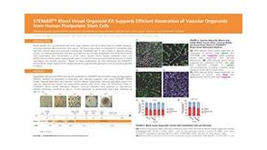 科学海报STEMdiff™ Blood Vessel Organoid Kit Supports Efficient Generation of Vascular Organoids from Human Pluripotent Stem Cells


 沪公网安备31010102008431号