Radujkovic A et al. ( )
Anticancer research 26 3A 2169--77
Combination treatment of imatinib-sensitive and -resistant BCR-ABL-positive CML cells with imatinib and farnesyltransferase inhibitors.
BACKGROUND: Resistance to imatinib monotherapy frequently emerges in advanced stages of chronic myelogenous leukemia (CML),supporting the rationale for combination drug therapy. In the present study,the activities of the farnesyltransferase inhibitors (FTIs) L744,832 and LB42918,as single agents and in combination with imatinib,were investigated in different imatinib-sensitive and -resistant BCR-ABL-positive CML cells. MATERIALS AND METHODS: Growth inhibition of the cell lines and primary patient cells was assessed by MTT assays and colony-forming cell assays,respectively. Drug interactions were analyzed according to the median-effect method of Chou and Talalay. The determination of apoptotic cell death was performed by annexin V/propidium iodide staining. RESULTS: Combinations of both FTIs with imatinib displayed synergism or sensitization (potentiation) in all the cell lines tested. In primary chronic phase CML cells,additive and synergistic effects were discernible for the combination of imatinib plus L744,832 and imatinib plus LB42918,respectively. Annexin V/propidium iodide staining showed enhancement of imatinib-induced apoptosis with either drug combination,both in imatinib-sensitive and -resistant cells. CONCLUSION: The results indicated the potential of L744,832 and LB42918 as combination agents for CML patients on imatinib treatment.
View Publication
产品号#:
84534
84544
产品名:
MethoCult GF H84534, 100mL
Tan W et al. (MAY 2006)
Journal of immunology (Baltimore,Md. : 1950) 176 10 6186--93
IL-17 receptor knockout mice have enhanced myelotoxicity and impaired hemopoietic recovery following gamma irradiation.
IL-17A is a T cell-derived proinflammatory cytokine required for microbial host defense. In vivo expression profoundly stimulates granulopoiesis. At baseline,the hemopoietic system of IL-17R knockout mice (IL-17Ra(-/-)) is,with the exception of increased splenic progenitor numbers,indistinguishable from normal control mice. However,when challenged with gamma irradiation,hemopoietic toxicity is significantly more pronounced in IL-17Ra(-/-) animals,with the gamma irradiation-associated LD(50) being reduced by 150 rad. In spleen-derived T cells,gamma irradiation induces significant murine IL-17A expression in vivo but not in vitro. After sublethal radiation injury (500 rad),the infusion of purified CD4(+) T cells enhances hemopoietic recovery. This recovery is significantly impaired in IL-17Ra(-/-) animals or after in vivo blockade of IL-17Ra in normal mice,resulting in a reduction of hemopoietic precursors by 50% and of neutrophils by 43%. Following sublethal radiation-induced myelosuppression,in vivo overexpression of murine IL-17A in normal mice substantially enhanced granulopoietic restoration in mice with a 4-fold increase in neutrophils and splenic precursors on day 8 (CFU-granulocyte-macrophage/granulocyte-erythrocyte-megakaryocyte-monocyte,CFU-high proliferative potential),as well as 2- and 3-fold increases of bone marrow precursors,respectively. This establishes IL-17A as a hemopoietic response cytokine to radiation injury in mice and an inducible mechanism that is required for recovery of granulopoiesis after radiation injury.
View Publication
Wendel H-G et al. (MAY 2006)
Proceedings of the National Academy of Sciences of the United States of America 103 19 7444--9
Loss of p53 impedes the antileukemic response to BCR-ABL inhibition.
Targeted cancer therapies exploit the continued dependence of cancer cells on oncogenic mutations. Such agents can have remarkable activity against some cancers,although antitumor responses are often heterogeneous,and resistance remains a clinical problem. To gain insight into factors that influence the action of a prototypical targeted drug,we studied the action of imatinib (STI-571,Gleevec) against murine cells and leukemias expressing BCR-ABL,an imatinib target and the initiating oncogene for human chronic myelogenous leukemia (CML). We show that the tumor suppressor p53 is selectively activated by imatinib in BCR-ABL-expressing cells as a result of BCR-ABL kinase inhibition. Inactivation of p53,which can accompany disease progression in human CML,impedes the response to imatinib in vitro and in vivo without preventing BCR-ABL kinase inhibition. Concordantly,p53 mutations are associated with progression to imatinib resistance in some human CMLs. Our results identify p53 as a determinant of the response to oncogene inhibition and suggest one way in which resistance to targeted therapy can emerge during the course of tumor evolution.
View Publication
Volpe DA and Warren MK (JUN 2003)
Toxicology in vitro : an international journal published in association with BIBRA 17 3 271--7
Myeloid clonogenic assays for comparison of the in vitro toxicity of alkylating agents.
A battery of clonal assays for myeloid progenitor cells (HPP-CFC,CFU-gemm,CFU-gm,CFU-g) was utilized to evaluate the myelotoxicity of a series of alkylating agents representing the spectrum of clinical times to nadir. Bone marrow aspirates from normal volunteers were incubated with mechlorethamine,busulfan,melphalan,carmustine or lomustine for 1 h and then cultured in methylcellulose with 30% serum and cytokines. There was a concentration-dependent inhibition of colony formation and often a differential toxicity to the myeloid progenitors with the alkylators tested. On a molar basis,mechlorethamine and melphalan were the most toxic of the alkylator drugs to the myeloid precursors. The most sensitive progenitor was CFU-gemm with the lowest inhibitory concentration IC(70) concentrations for mechlorethamine,melphalan,carmustine and lomustine. Generally,there was great similarity for drug effects between CFU-g and CFU-gm with overlapping inhibition curves. HPP-CFC proved to be the least sensitive of the progenitors to the toxic actions of the drugs. While there was no correlation between the time to clinical neutropenic nadir and the most sensitive progenitor in the clonal assays,the CFU-gm assay remains a suitable method for determining the myelotoxic potential of cytotoxic agents.
View Publication
Jones DC et al. (JUL 2003)
Journal of immunology 171 1 196--203
Peroxisome proliferator-activated receptor alpha negatively regulates T-bet transcription through suppression of p38 mitogen-activated protein kinase activation.
Expression of the nuclear hormone receptor peroxisome proliferator-activated receptor alpha (PPARalpha) in resting lymphocytes was recently established,although the physiologic role(s) played by this nuclear hormone receptor in these cell types remains unresolved. In this study,we used CD4(+) T cells isolated from PPARalpha(-/-) and wild-type mice,as well as cell lines that constitutively express PPARalpha,in experiments designed to evaluate the role of this hormone receptor in the regulation of T cell function. We report that activated CD4(+) T cells lacking PPARalpha produce increased levels of IFN-gamma,but significantly lower levels of IL-2 when compared with activated wild-type CD4(+) T cells. Furthermore,we demonstrate that PPARalpha regulates the expression of these cytokines by CD4(+) T cells in part,through its ability to negatively regulate the transcription of T-bet. The induction of T-bet expression in CD4(+) T cells was determined to be positively influenced by p38 mitogen-activated protein (MAP) kinase activation,and the presence of unliganded PPARalpha effectively suppressed the phosphorylation of p38 MAP kinase. The activation of PPARalpha with highly specific ligands relaxed its capacity to suppress p38 MAP kinase phosphorylation and promoted T-bet expression. These results demonstrate a novel DNA-binding independent and agonist-controlled regulatory influence by the nuclear hormone receptor PPARalpha.
View Publication

 EasySep™小鼠TIL(CD45)正选试剂盒


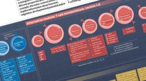 挂图T Cell Nomenclature: From Subsets to Modules A modular framework for classifying T cells by lineage, function, migration, differentiation, and antigen context.
 实验方案Optimizing Delivery Efficiency with Fluorescent Dextran Using the CellPore™ Transfection System


 沪公网安备31010102008431号