S. Ihara et al. (JUN 2018)
Journal of Crohn's & colitis
Adhesive interactions between Mononuclear Phagocytes and Intestinal Epithelium Perturb Normal Epithelial Differentiation and Serve as a Therapeutic Target in Inflammatory Bowel Disease.
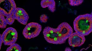
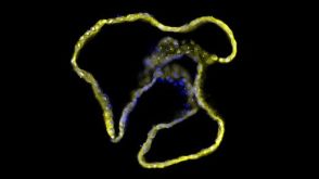
Background and Aims Disturbance of intestinal homeostasis is associated with the development of inflammatory bowel disease (IBD),and TGF-beta$ signaling impairment in mononuclear phagocytes (MPs) causes murine colitis with goblet cell depletion. Here,we examined an organoid-MP co-culture system to study the role of MPs in intestinal epithelial differentiation and homeostasis. Methods Intestinal organoids were co-cultured with lamina propria leukocytes and bone marrow-derived dendritic cells (BMDCs) from CD11c-cre Tgfbr2fl/fl mice. Organoid-MP adhesive interactions were evaluated by microscopy,RT-PCR,and flow cytometry. Murine colitis models (dextran sodium sulphate (DSS),CD11c-cre Tgfbr2fl/fl,T-cell-transfer) were used for histological and immunohistochemical analysis. Anti-E-cadherin antibody treatment or CD11c+-cell-specific CDH1 gene deletion were performed for E-cadherin neutralization or knockout. Colonic biopsies from patients with ulcerative colitis were analyzed by flow cytometry. Results Intestinal organoids co-cultured with CD11c+ lamina propria leukocytes or BMDCs from CD11c-cre Tgfbr2fl/fl mice showed morphological changes and goblet cell depletion with Notch signal activation,analogous to CD11c-cre Tgfbr2fl/fl colitis. E-cadherin was upregulated in CD11c+ MPs,especially CX3CR1+CCR2+ monocytes,of CD11c-cre Tgfbr2fl/fl mice. E-cadherin-mediated BMDC adhesion promoted Notch activation and cystic changes in organoids. Anti-E-cadherin antibody treatment attenuated colitis in CD11c-cre Tgfbr2fl/fl and T-cell-transferred mice. In addition,E-cadherin deletion in CD11c+ cells attenuated colitis in both CD11c-cre Tgfbr2fl/fl and DSS-treated mice. In patients with ulcerative colitis,E-cadherin expressed by intestinal CD11c+ leukocytes was enhanced compared with that in healthy controls. Conclusions E-cadherin-mediated MP-epithelium adhesion is associated with the development of colitis,and blocking these adhesions may have therapeutic potential for IBD.
View Publication
产品号#:
06005
产品名:
IntestiCult™ 类器官生长培养基 (小鼠)
Qiao Y et al. (APR 2011)
Cancer research 71 8 3076--86
FOXQ1 regulates epithelial-mesenchymal transition in human cancers.
Epithelial-mesenchymal transition (EMT) in cancer cells plays a pivotal role in determining metastatic prowess,but knowledge of EMT regulation remains incomplete. In this study,we defined a critical functional role for the Forkhead transcription factor FOXQ1 in regulating EMT in breast cancer cells. FOXQ1 expression was correlated with high-grade basal-like breast cancers and was associated with poor clinical outcomes. RNAi-mediated suppression of FOXQ1 expression in highly invasive human breast cancer cells reversed EMT,reduced invasive ability,and alleviated other aggressive cancer phenotypes manifested in 3-dimensional Matrigel (BD Biosciences) culture. Conversely,enforced expression of FOXQ1 in differentiated human mammary epithelial cells (HMLER) or epithelial cancer cell lines provoked an epithelial to mesenchymal morphologic change,gain of stem cell-like properties,and acquisition of resistance to chemotherapy-induced apoptosis. Mechanistic investigations revealed that FOXQ1-induced EMT was associated with transcriptional inactivation of the epithelial regulator E-cadherin (CDH1). Our findings define a key role for FOXQ1 in regulating EMT and aggressiveness in human cancer.
View Publication
Wang H et al. (JAN 2012)
Journal of translational medicine 10 1 167
Oncolytic vaccinia virus GLV-1h68 strain shows enhanced replication in human breast cancer stem-like cells in comparison to breast cancer cells.
BACKGROUND: Recent data suggest that cancer stem cells (CSCs) play an important role in cancer,as these cells possess enhanced tumor-forming capabilities and are responsible for relapses after apparently curative therapies have been undertaken. Hence,novel cancer therapies will be needed to test for both tumor regression and CSC targeting. The use of oncolytic vaccinia virus (VACV) represents an attractive anti-tumor approach and is currently under evaluation in clinical trials. The purpose of this study was to demonstrate whether VACV does kill CSCs that are resistant to irradiation and chemotherapy. METHODS: Cancer stem-like cells were identified and separated from the human breast cancer cell line GI-101A by virtue of increased aldehyde dehydrogenase 1 (ALDH1) activity as assessed by the ALDEFLUOR assay and cancer stem cell-like features such as chemo-resistance,irradiation-resistance and tumor-initiating were confirmed in cell culture and in animal models. VACV treatments were applied to both ALDEFLUOR-positive cells in cell culture and in xenograft tumors derived from these cells. Moreover,we identified and isolated CD44(+)CD24(+)ESA(+) cells from GI-101A upon an epithelial-mesenchymal transition (EMT). These cells were similarly characterized both in cell culture and in animal models. RESULTS: We demonstrated for the first time that the oncolytic VACV GLV-1h68 strain replicated more efficiently in cells with higher ALDH1 activity that possessed stem cell-like features than in cells with lower ALDH1 activity. GLV-1h68 selectively colonized and eventually eradicated xenograft tumors originating from cells with higher ALDH1 activity. Furthermore,GLV-1h68 also showed preferential replication in CD44(+)CD24(+)ESA(+) cells derived from GI-101A upon an EMT induction as well as in xenograft tumors originating from these cells that were more tumorigenic than CD44(+)CD24(-)ESA(+) cells. CONCLUSIONS: Taken together,our findings indicate that GLV-1h68 efficiently replicates and kills cancer stem-like cells. Thus,GLV-1h68 may become a promising agent for eradicating both primary and metastatic tumors,especially tumors harboring cancer stem-like cells that are resistant to chemo and/or radiotherapy and may be responsible for recurrence of tumors.
View Publication
产品号#:
01700
01705
05620
01702
产品名:
ALDEFLUOR™ 试剂盒
ALDEFLUOR™ DEAB试剂, 1.5 mM, 1 mL
MammoCult™ 人源培养基套装
ALDEFLUOR™检测缓冲液
Wang Y et al. (MAR 2017)
Mucosal immunology 10 2 373--384
An LGG-derived protein promotes IgA production through upregulation of APRIL expression in intestinal epithelial cells.
p40,a Lactobacillus rhamnosus GG (LGG)-derived protein,transactivates epidermal growth factor receptor (EGFR) in intestinal epithelial cells,leading to amelioration of intestinal injury and inflammation. To elucidate mechanisms by which p40 regulates mucosal immunity to prevent inflammation,this study aimed to determine the effects and mechanisms of p40 on regulation of a proliferation-inducing ligand (APRIL) expression in intestinal epithelial cells for promoting immunoglobulin A (IgA) production. p40 upregulated April gene expression and protein production in mouse small intestine epithelial (MSIE) cells,which were inhibited by blocking EGFR expression and kinase activity. Enteroids from Egfr(fl/fl),but not Egfr(fl/fl)-Vil-Cre mice with EGFR specifically deleted in intestinal epithelial cells,exhibited increased April gene expression by p40 treatment. p40-conditioned media from MSIE cells increased B-cell class switching to IgA(+) cells and IgA production,which was suppressed by APRIL receptor-neutralizing antibodies. Treatment of B cells with p40 did not show any effects on IgA production. p40 treatment increased April gene expression and protein production in small intestinal epithelial cells,fecal IgA levels,IgA(+)B220(+),IgA(+)CD19(+),and IgA(+) plasma cells in lamina propria of Egfr(fl/fl),but not of Egfr(fl/fl)-Vil-Cre,mice. Thus p40 upregulates EGFR-dependent APRIL production in intestinal epithelial cells,which may contribute to promoting IgA production.
View Publication
产品号#:
06005
产品名:
IntestiCult™ 类器官生长培养基 (小鼠)
Ibiza S et al. (JUL 2016)
Nature 535 7612 440--443
Glial-cell-derived neuroregulators control type 3 innate lymphoid cells and gut defence.
Group 3 innate lymphoid cells (ILC3) are major regulators of inflammation and infection at mucosal barriers. ILC3 development is thought to be programmed,but how ILC3 perceive,integrate and respond to local environmental signals remains unclear. Here we show that ILC3 in mice sense their environment and control gut defence as part of a glial"ILC3"epithelial cell unit orchestrated by neurotrophic factors. We found that enteric ILC3 express the neuroregulatory receptor RET. ILC3-autonomous Ret ablation led to decreased innate interleukin-22 (IL-22),impaired epithelial reactivity,dysbiosis and increased susceptibility to bowel inflammation and infection. Neurotrophic factors directly controlled innate Il22 downstream of the p38 MAPK/ERK-AKT cascade and STAT3 activation. Notably,ILC3 were adjacent to neurotrophic-factor-expressing glial cells that exhibited stellate-shaped projections into ILC3 aggregates. Glial cells sensed microenvironmental cues in a MYD88-dependent manner to control neurotrophic factors and innate IL-22. Accordingly,glial-intrinsic Myd88 deletion led to impaired production of ILC3-derived IL-22 and a pronounced propensity towards gut inflammation and infection. Our work sheds light on a novel multi-tissue defence unit,revealing that glial cells are central hubs of neuron and innate immune regulation by neurotrophic factor signals.
View Publication
产品号#:
06005
产品名:
IntestiCult™ 类器官生长培养基 (小鼠)
Saxena A et al. (JUL 2017)
American journal of physiology. Gastrointestinal and liver physiology 313 1 G26--G38
Absence of the NOD2 protein renders epithelia more susceptible to barrier dysfunction due to mitochondrial dysfunction.
Irregular mitochondria structure and reduced ATP in some patients with IBD suggest that metabolic stress contributes to disease. Loss-of-function mutation in the nucleotide-binding oligomerization domain (NOD)-2 gene is a major susceptibility trait for IBD. Hence,we assessed if loss of NOD2 further impairs the epithelial barrier function instigated by disruption of mitochondrial ATP synthesis via the hydrogen ionophore dinitrophenol (DNP). NOD2 protein (virtually undetectable in epithelia under basal conditions) was increased in T84 (human colon cell line) cells treated with noninvasive Escherichia coli + DNP (16 h). Increased intracellular bacteria in wild-type (WT) and NOD2 knockdown (KD) cells and colonoids from NOD2(-/-) mice were mediated by reactive oxygen species (ROS) and the MAPK ERK1/2 pathways as determined by cotreatment with the antioxidant mitoTEMPO and the ERK inhibitor U0126: ROS was upstream of ERK1/2 activation. Despite increased E. coli in DNP-treated NOD2 KD compared with WT cells,there were no differences in the internalization of fluorescent inert beads or dead E. coli particles. This suggests that lack of killing in the NOD2 KD cells was responsible for the increased numbers of viable intracellular bacteria; a conclusion supported by evidence of reduced autophagy in NOD2 KD T84 epithelia. Thus,in a two-hit hypothesis,decreased barrier function due to dysfunctional mitochondrial is amplified by lack of NOD2 in transporting enterocytes: subsequently,greater numbers of bacteria entering the mucosa would be a significant inflammatory threat especially since individuals with NOD2 mutations have compromised macrophage and Paneth cell responses to bacteria.NEW & NOTEWORTHY Increased internalization of bacteria by epithelia with dysfunctional mitochondria (reduced ATP) is potentiated if the cells lack nucleotide-binding oligomerization domain 2 (NOD2),mutations in which are inflammatory bowel disease-susceptibility traits. Uptake of bacteria was dependent on reactive oxygen species and MAP-kinase activity,and the increased viable intracellular bacteria in NOD2(-/-) cells likely reflect a reduced ability to recognize and kill bacteria. Thus a significant barrier defect occurs with NOD2 deficiency in conjunction with metabolic stress that could contribute to inflammation.
View Publication

 EasySep™小鼠TIL(CD45)正选试剂盒


 沪公网安备31010102008431号