
 EasySep™小鼠TIL(CD45)正选试剂盒


技术资料
-
 科学海报A Feeder-Free and Serum-Free Culture System Supports Expansion of CD34+ AML and CML Stem/Progenitor Cells
Conference:
ISEH 2019
-

-
-
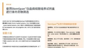
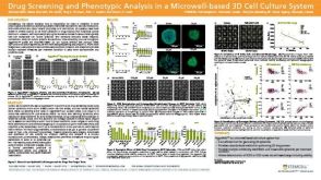 科学海报Drug Screening and Phenotypic Analysis in a Microwell-based 3D Cell Culture System
Conference:
AACR 2018


 沪公网安备31010102008431号