Heberden C et al. (NOV 2013)
The Journal of Steroid Biochemistry and Molecular Biology 138 395--402
Dexamethasone inhibits the maturation of newly formed neurons and glia supplemented with polyunsaturated fatty acids
Stress bears a negative impact on adult neurogenesis. High levels of corticoids have been shown to inhibit neural stem cell proliferation,and are considered responsible for the loss of neural precursors. Their effects on the differentiation of the glial and neuronal lineages have been less studied. We examined the effect of dexamethasone (Dex),a synthetic glucocorticoid,on the differentiation of rat neural stem cells in vitro. Dex had no effect on the differentiation of cells cultured under standard conditions. Since we previously determined that NSC,when cultured under classical conditions,were deprived of polyunsaturated fatty acids (PUFA),and displayed phospholipid compositions very different from the in vivo figures [1],we examined the effect of Dex under PUFA supplementation. Dex impaired neuron and oligodendrocyte maturation in PUFA-supplemented cells,demonstrated by the reduction of neurite lengths and oligodendrocyte sizes. This effect was mediated by the glucocorticoid receptor (GR),since it was eliminated by mifepristone,a GR antagonist,and could be relayed by a reduction of ERK phosphorylation. We determined that GR was associated with PPAR β and α under basal conditions,and that this association was disrupted when PUFA were added in combination with Dex. We assumed that this effect on the receptor status enabled the effect of Dex on PUFA supplemented cells,since we determined that the binding to the glucocorticoid response element was higher in cells incubated with PUFA and Dex. In conclusion,corticoids can impair NSC differentiation,and consequently impact the entire process of neurogenesis.
View Publication
产品号#:
05771
产品名:
Hjelm BE et al. (SEP 2013)
Human Molecular Genetics 22 17 3534--3546
In vitro-differentiated neural cell cultures progress towards donor-identical brain tissue
Multiple research groups have observed neuropathological phenotypes and molecular symptoms in vitro using induced pluripotent stem cell (iPSC)-derived neural cell cultures (i.e. patient-specific neurons and glia). However,the global differences/similarities that may exist between in vitro neural cells and their tissue-derived counterparts remain largely unknown. In this study,we compared temporal series of iPSC-derived in vitro neural cell cultures to endogenous brain tissue from the same autopsy donor. Specifically,we utilized RNA sequencing (RNA-Seq) to evaluate the transcriptional progression of in vitro-differentiated neural cells (over a timecourse of 0,35,70,105 and 140 days),and compared this with donor-identical temporal lobe tissue. We observed in vitro progression towards the reference brain tissue,and the following three results support this conclusion: (i) there was a significant increasing monotonic correlation between the days of our timecourse and the number of actively transcribed protein-coding genes and long intergenic non-coding RNAs (lincRNAs) (P < 0.05),consistent with the transcriptional complexity of the brain; (ii) there was an increase in CpG methylation after neural differentiation that resembled the epigenomic signature of the endogenous tissue; and (iii) there was a significant decreasing monotonic correlation between the days of our timecourse and the percent of in vitro to brain-tissue differences (P < 0.05) for tissue-specific protein-coding genes and all putative lincRNAs. Taken together,these results are consistent with in vitro neural development and physiological progression occurring predominantly by transcriptional activation of downregulated genes rather than deactivation of upregulated genes.
View Publication
产品号#:
05750
05751
05752
产品名:
NeuroCult™ NS-A 基础培养基(人)
NeuroCult™ NS-A 扩增试剂盒(人)
NeuroCult™ NS-A 分化试剂盒(人)
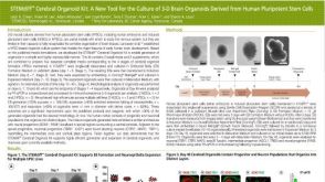
K. B. Langer et al. (APR 2018)
Stem cell reports 10 4 1282--1293
Retinal Ganglion Cell Diversity and Subtype Specification from Human Pluripotent Stem Cells.
Retinal ganglion cells (RGCs) are the projection neurons of the retina and transmit visual information to postsynaptic targets in the brain. While this function is shared among nearly all RGCs,this class of cell is remarkably diverse,comprised of multiple subtypes. Previous efforts have identified numerous RGC subtypes in animal models,but less attention has been paid to human RGCs. Thus,efforts of this study examined the diversity of RGCs differentiated from human pluripotent stem cells (hPSCs) and characterized defined subtypes through the expression of subtype-specific markers. Further investigation of these subtypes was achieved using single-cell transcriptomics,confirming the combinatorial expression of molecular markers associated with these subtypes,and also provided insight into more subtype-specific markers. Thus,the results of this study describe the derivation of RGC subtypes from hPSCs and will support the future exploration of phenotypic and functional diversity within human RGCs.
View Publication
产品号#:
05790
05792
05793
05794
05795
85850
85857
产品名:
BrainPhys™神经元培养基
BrainPhys™神经元培养基和SM1试剂盒
BrainPhys™ 神经元培养基N2-A和SM1试剂盒
BrainPhys™原代神经元试剂盒
BrainPhys™ hPSC 神经元试剂盒
mTeSR™1
mTeSR™1
A. M. Tukker et al. (JUL 2018)
Neurotoxicology 67 215--225
Human iPSC-derived neuronal models for in vitro neurotoxicity assessment.
Neurotoxicity testing still relies on ethically debated,expensive and time consuming in vivo experiments,which are unsuitable for high-throughput toxicity screening. There is thus a clear need for a rapid in vitro screening strategy that is preferably based on human-derived neurons to circumvent interspecies translation. Recent availability of commercially obtainable human induced pluripotent stem cell (hiPSC)-derived neurons and astrocytes holds great promise in assisting the transition from the current standard of rat primary cortical cultures to an animal-free alternative. We therefore composed several hiPSC-derived neuronal models with different ratios of excitatory and inhibitory neurons in the presence or absence of astrocytes. Using immunofluorescent stainings and multi-well micro-electrode array (mwMEA) recordings we demonstrate that these models form functional neuronal networks that become spontaneously active. The differences in development of spontaneous neuronal activity and bursting behavior as well as spiking patterns between our models confirm the importance of the presence of astrocytes. Preliminary neurotoxicity assessment demonstrates that these cultures can be modulated with known seizurogenic compounds,such as picrotoxin (PTX) and endosulfan,and the neurotoxicant methylmercury (MeHg). However,the chemical-induced effects on different parameters for neuronal activity,such as mean spike rate (MSR) and mean burst rate (MBR),may depend on the ratio of inhibitory and excitatory neurons. Our results thus indicate that hiPSC-derived neuronal models must be carefully designed and characterized prior to large-scale use in neurotoxicity screening.
View Publication
Binder LI et al. (SEP 1984)
Proceedings of the National Academy of Sciences of the United States of America 81 17 5613--7
Heterogeneity of microtubule-associated protein 2 during rat brain development.
The electrophoretic pattern of the large microtubule-associated protein,MAP2,changes during rat brain development. Immunoblots of NaDodSO4 extracts obtained from the cerebral cortex,cerebellum,and thalamus at 10-15 days after birth reveal only a single electrophoretic species when probed with any of three MAP2 monoclonal antibodies. By contrast,adult MAP2 contains two immunoreactive species,MAP2a and MAP2b. The single band of MAP2 from immature brain electrophoretically comigrates with adult MAP2b. Between postnatal days 17 and 18,immature MAP2 simultaneously resolves into two species in both the cerebellum and cerebral cortex. Immunoblots of NaDodSO4 extracts from spinal cord demonstrate the adult complement of MAP2 by day 10,indicating that MAP2 does not change coordinately throughout the entire central nervous system. In vitro cAMP-dependent phosphorylation of immature MAP2 causes a band split reminiscent of that seen during brain development in vivo. The possibility that the developmentally regulated changes observed in MAP2 during brain maturation are due to timed phosphorylation events is discussed.
View Publication
Kishigami S et al. (FEB 2006)
Biochemical and biophysical research communications 340 1 183--9
Significant improvement of mouse cloning technique by treatment with trichostatin A after somatic nuclear transfer.
The low success rate of animal cloning by somatic cell nuclear transfer (SCNT) is believed to be associated with epigenetic errors including abnormal DNA hypermethylation. Recently,we elucidated by using round spermatids that,after nuclear transfer,treatment of zygotes with trichostatin A (TSA),an inhibitor of histone deacetylase,can remarkably reduce abnormal DNA hypermethylation depending on the origins of transferred nuclei and their genomic regions [S. Kishigami,N. Van Thuan,T. Hikichi,H. Ohta,S. Wakayama. E. Mizutani,T. Wakayama,Epigenetic abnormalities of the mouse paternal zygotic genome associated with microinsemination of round spermatids,Dev. Biol. (2005) in press]. Here,we found that 5-50 nM TSA-treatment for 10 h following oocyte activation resulted in more efficient in vitro development of somatic cloned embryos to the blastocyst stage from 2- to 5-fold depending on the donor cells including tail tip cells,spleen cells,neural stem cells,and cumulus cells. This TSA-treatment also led to more than 5-fold increase in success rate of mouse cloning from cumulus cells without obvious abnormality but failed to improve ES cloning success. Further,we succeeded in establishment of nuclear transfer-embryonic stem (NT-ES) cells from TSA-treated cloned blastocyst at a rate three times higher than those from untreated cloned blastocysts. Thus,our data indicate that TSA-treatment after SCNT in mice can dramatically improve the practical application of current cloning techniques.
View Publication
产品号#:
05700
05701
05702
72282
72284
产品名:
NeuroCult™ 基础培养基(小鼠和大鼠)
NeuroCult™ 扩增添加物(小鼠和大鼠)
NeuroCult™扩增试剂盒(小鼠和大鼠)
曲古抑菌素 A(Trichostatin A)
曲古抑菌素 A(Trichostatin A)
Zhang Z et al. (JAN 2006)
Human molecular genetics 15 2 337--46
Palmitoyl-protein thioesterase-1 deficiency mediates the activation of the unfolded protein response and neuronal apoptosis in INCL.
Numerous proteins undergo modification by palmitic acid (S-acylation) for their biological functions including signal transduction,vesicular transport and maintenance of cellular architecture. Although palmitoylation is an essential modification,these proteins must also undergo depalmitoylation for their degradation by lysosomal proteases. Palmitoyl-protein thioesterase-1 (PPT1),a lysosomal enzyme,cleaves thioester linkages in S-acylated proteins and removes palmitate residues facilitating the degradation of these proteins. Thus,inactivating mutations in the PPT1 gene cause infantile neuronal ceroid lipofuscinosis (INCL),a devastating neurodegenerative storage disorder of childhood. Although rapidly progressing brain atrophy is the most dramatic pathological manifestation of INCL,the molecular mechanism(s) remains unclear. Using PPT1-knockout (PPT1-KO) mice that mimic human INCL,we report here that the endoplasmic reticulum (ER) in the brain cells of these mice is structurally abnormal. Further,we demonstrate that the level of growth-associated protein-43 (GAP-43),a palmitoylated neuronal protein,is elevated in the brains of PPT1-KO mice. Moreover,forced expression of GAP-43 in PPT1-deficient cells results in the abnormal accumulation of this protein in the ER. Consistent with these results,we found evidence for the activation of unfolded protein response (UPR) marked by elevated levels of phosphorylated translation initiation factor,eIF2alpha,increased expression of chaperone proteins such as glucose-regulated protein-78 and activation of caspase-12,a cysteine proteinase in the ER,mediating caspase-3 activation and apoptosis. Our results,for the first time,link PPT1 deficiency with the activation of UPR,apoptosis and neurodegeneration in INCL and identify potential targets for therapeutic intervention in this uniformly fatal disease.
View Publication
产品号#:
05700
05701
05702
产品名:
NeuroCult™ 基础培养基(小鼠和大鼠)
NeuroCult™ 扩增添加物(小鼠和大鼠)
NeuroCult™扩增试剂盒(小鼠和大鼠)
Li J-M et al. (FEB 2007)
Molecular endocrinology (Baltimore,Md.) 21 2 499--511
Angiotensin II-induced neural differentiation via angiotensin II type 2 (AT2) receptor-MMS2 cascade involving interaction between AT2 receptor-interacting protein and Src homology 2 domain-containing protein-tyrosine phosphatase 1.
Angiotensin II (Ang II) type 2 (AT2) receptors are abundantly expressed not only in the fetal brain where they probably contribute to brain development,but also in pathological conditions to protect the brain against stroke; however,the detailed mechanisms are unclear. Here,we demonstrated that AT2 receptor signaling induced neural differentiation via an increase in MMS2,one of the ubiquitin-conjugating enzyme variants. The AT2 receptor,MMS2,Src homology 2 domain-containing protein-tyrosine phosphatase 1 (SHP-1),and newly cloned AT2 receptor-interacting protein (ATIP) were highly expressed in fetal rat neurons and declined after birth. Ang II induced MMS2 expression in a dose-dependent manner,reaching a peak after 4 h of stimulation,and this effect was enhanced with AT1 receptor blocker,valsartan,but inhibited by AT2 receptor blocker PD123319. Moreover,we observed that an AT2 receptor agonist,CGP42112A,alone enhanced MMS2 expression. Neurons treated with small interfering RNA of MMS2 failed to exhibit neurite outgrowth and synapse formation. Moreover,the increase in AT2 receptor-induced MMS2 mRNA expression was enhanced by overexpression of ATIP but inhibited by small interfering RNA of SHP-1 and overexpression of catalytically dominant-negative SHP-1 or a tyrosine phosphatase inhibitor,sodium orthovanadate. After AT2 receptor stimulation,ATIP and SHP-1 were translocated into the nucleus after formation of their complex. Furthermore,increased MMS2 expression mediates the inhibitor of DNA binding 1 proteolysis and promotes DNA repair. These results provide a new insight into the contribution of AT2 receptor stimulation to neural differentiation via transactivation of MMS2 expression involving the association of ATIP and SHP-1.
View Publication
产品号#:
05700
05703
05704
产品名:
NeuroCult™ 基础培养基(小鼠和大鼠)
NeuroCult™ 分化添加物(小鼠和大鼠)
NeuroCult™ 分化试剂盒(小鼠和大鼠)
Walker TL et al. (APR 2007)
The Journal of neuroscience : the official journal of the Society for Neuroscience 27 14 3734--42
The doublecortin-expressing population in the developing and adult brain contains multipotential precursors in addition to neuronal-lineage cells.
Doublecortin (DCX) has recently been promulgated as a selective marker of cells committed to the neuronal lineage in both the developing and the adult brain. To explore the potential of DCX-positive (DCX+) cells more stringently,these cells were isolated by flow cytometry from the brains of transgenic mice expressing green fluorescent protein under the control of the DCX promoter in embryonic,early postnatal,and adult animals. It was found that virtually all of the cells (99.9%) expressing high levels of DCX (DCX(high)) in the embryonic brain coexpressed the neuronal marker betaIII-tubulin and that this population contained no stem-like cells as demonstrated by lack of neurosphere formation in vitro. However,the DCX+ population from the early postnatal brain and the adult subventricular zone and hippocampus,which expressed low levels of DCX (DCX(low)),was enriched for neurosphere-forming cells,with only a small subpopulation of these cells coexpressing the neuronal markers betaIII-tubulin or microtubule-associated protein 2. Similarly,the DCX(low) population from embryonic day 14 (E14) brain contained neurosphere-forming cells. Only the postnatal cerebellum and adult olfactory bulb contained some DCX(high) cells,which were shown to be similar to the E14 DCX(high) cells in that they had no stem cell activity. Electrophysiological studies confirmed the heterogeneous nature of DCX+ cells,with some cells displaying characteristics of immature or mature neurons,whereas others showed no neuronal characteristics whatsoever. These results indicate that DCX(high) cells,regardless of location,are restricted to the neuronal lineage or are bone fide neurons,whereas some DCX(low) cells retain their multipotentiality.
View Publication

 EasySep™小鼠TIL(CD45)正选试剂盒


 沪公网安备31010102008431号