Shahbazi M et al. (JUL 2013)
Journal of the Neurological Sciences 330 1–2 85--93
Inhibitory effects of neural stem cells derived from human embryonic stem cells on differentiation and function of monocyte-derived dendritic cells
Neural stem cells (NSCs) possess immunosuppressive characteristics,but effects of NSCs on human dendritic cells (DCs),the most important antigen presenting cells,are less well studied. We used an in vitro approach to evaluate the effects of human NSCs on differentiation of human blood CD14+ monocytes into DCs. NSCs derived from H1 human embryonic stem cells (hESC-NSCs) and human ReNcell NSC line,as well as human bone marrow derived mesenchymal stem cells (MSCs),were tested. We observed that in response to treatment with interleukin-4 and granulocyte macrophage colony-stimulating factor CD14+ monocytes co-cultured with NSCs were able to down-regulate CD14 and up-regulate the differentiation marker CD1a,whereas MSC co-culture strongly inhibited CD1a expression and supported prolonged expression of CD14. A similar difference between NSCs and MSCs was noted when lipopolysaccharides were included to induce maturation of monocyte-derived DCs. However,when effects on the function of derived DCs were investigated,NSCs suppressed the elevation of the DC maturation marker CD83,although not the up-regulation of costimulatory molecules CD80,CD86 and CD40,and impaired the functional capacity of the derived DCs to stimulate alloreactive T cells. We did not observe any obvious difference between hESC-NSCs and ReNcell NSCs in inhibiting DC maturation and function. Our data suggest that although human NSCs are less effective than human MSCs in suppressing monocyte differentiation into DCs,these stem cells can still affect the function of DCs,ultimately regulating specific immune responses.
View Publication
产品号#:
05850
05857
05870
05875
85850
85857
85870
85875
70025
70025.1
70025.2
70025.3
70047
70047.1
70047.2
70048
70048.1
70048.2
产品名:
mTeSR™1
mTeSR™1
冻存的人外周血单个核细胞
冻存的人外周血单个核细胞
冻存的人外周血单个核细胞
冻存的人外周血单个核细胞
Conte D et al. (JAN 2012)
PloS one 7 12 e52167
Loss of Atrx sensitizes cells to DNA damaging agents through p53-mediated death pathways.
Prevalent cell death in forebrain- and Sertoli cell-specific Atrx knockout mice suggest that Atrx is important for cell survival. However,conditional ablation in other tissues is not associated with increased death indicating that diverse cell types respond differently to the loss of this chromatin remodeling protein. Here,primary macrophages isolated from Atrx(f/f) mice were infected with adenovirus expressing Cre recombinase or β-galactosidase,and assayed for cell survival under different experimental conditions. Macrophages survive without Atrx but undergo rapid apoptosis upon lipopolysaccharide (LPS) activation suggesting that chromatin reorganization in response to external stimuli is compromised. Using this system we next tested the effect of different apoptotic stimuli on cell survival. We observed that survival of Atrx-null cells were similar to wild type cells in response to serum withdrawal,anti-Fas antibody,C2 ceramide or dexamethasone treatment but were more sensitive to 5-fluorouracil (5-FU). Cell survival could be rescued by re-introducing Atrx or by removal of p53 demonstrating the cell autonomous nature of the effect and its p53-dependence. Finally,we demonstrate that multiple primary cell types (myoblasts,embryonic fibroblasts and neurospheres) were sensitive to 5-FU,cisplatin,and UV light treatment. Together,our results suggest that cells lacking Atrx are more sensitive to DNA damaging agents and that this may result in enhanced death during development when cells are at their proliferative peak. Moreover,it identifies potential treatment options for cancers associated with ATRX mutations,including glioblastoma and pancreatic neuroendocrine tumors.
View Publication
产品号#:
05700
05701
05702
产品名:
NeuroCult™ 基础培养基(小鼠和大鼠)
NeuroCult™ 扩增添加物(小鼠和大鼠)
NeuroCult™扩增试剂盒(小鼠和大鼠)
Binda E et al. (DEC 2012)
Cancer cell 22 6 765--80
The EphA2 receptor drives self-renewal and tumorigenicity in stem-like tumor-propagating cells from human glioblastomas.
In human glioblastomas (hGBMs),tumor-propagating cells with stem-like characteristics (TPCs) represent a key therapeutic target. We found that the EphA2 receptor tyrosine kinase is overexpressed in hGBM TPCs. Cytofluorimetric sorting into EphA2(High) and EphA2(Low) populations demonstrated that EphA2 expression correlates with the size and tumor-propagating ability of the TPC pool in hGBMs. Both ephrinA1-Fc,which caused EphA2 downregulation in TPCs,and siRNA-mediated knockdown of EPHA2 expression suppressed TPCs self-renewal ex vivo and intracranial tumorigenicity,pointing to EphA2 downregulation as a causal event in the loss of TPCs tumorigenicity. Infusion of ephrinA1-Fc into intracranial xenografts elicited strong tumor-suppressing effects,suggestive of therapeutic applications.
View Publication
产品号#:
05751
产品名:
NeuroCult™ NS-A 扩增试剂盒(人)
Maynard KR and Stein E (NOV 2012)
The Journal of neuroscience : the official journal of the Society for Neuroscience 32 47 16637--50
DSCAM contributes to dendrite arborization and spine formation in the developing cerebral cortex.
Down syndrome cell adhesion molecule,or DSCAM,has been implicated in many neurodevelopmental processes including axon guidance,dendrite arborization,and synapse formation. Here we show that DSCAM plays an important role in regulating the morphogenesis of cortical pyramidal neurons in the mouse. We report that DSCAM expression is developmentally regulated and localizes to synaptic plasma membranes during a time of robust cortical dendrite arborization and spine formation. Analysis of mice that carry a spontaneous mutation in DSCAM (DSCAM(del17)) revealed gross morphological changes in brain size and shape in addition to subtle changes in cortical organization,volume,and lamination. Early postnatal mutant mice displayed a transient decrease in cortical thickness,but these reductions could not be attributed to changes in neuron production or cell death. DSCAM(del17) mutants showed temporary impairments in the branching of layer V pyramidal neuron dendrites at P10 and P17 that recovered to normal by adulthood. Defects in DSCAM(del17) dendrite branching correlated with a temporal increase in apical branch spine density and lasting changes in spine morphology. At P15 and P42,mutant mice displayed a decrease in the percentage of large,stable spines and an increase in the percentage of small,immature spines. Together,our findings suggest that DSCAM contributes to pyramidal neuron morphogenesis by regulating dendrite arborization and spine formation during cortical circuit development.
View Publication
产品号#:
05715
产品名:
NeuroCult™成年中枢神经系统(CNS)组织酶解试剂盒(小鼠和大鼠)
Gerardo Valadez J et al. (JAN 2013)
Cancer letters 328 2 297--306
Identification of Hedgehog pathway responsive glioblastomas by isocitrate dehydrogenase mutation.
The Hedgehog (Hh) pathway regulates the growth of a subset of adult gliomas and better definition of Hh-responsive subtypes could enhance the clinical utility of monitoring and targeting this pathway in patients. Somatic mutations of the isocitrate dehydrogenase (IDH) genes occur frequently in WHO grades II and III gliomas and WHO grade IV secondary glioblastomas. Hh pathway activation in WHO grades II and III gliomas suggests that it might also be operational in glioblastomas that developed from lower-grade lesions. To evaluate this possibility and to better define the molecular and histopathological glioma subtypes that are Hh-responsive,IDH genes were sequenced in adult glioma specimens assayed for an operant Hh pathway. The proportions of grades II-IV specimens with IDH mutations correlated with the proportions that expressed elevated levels of the Hh gene target PTCH1. Indices of an operational Hh pathway were measured in all primary cultures and xenografts derived from IDH-mutant glioma specimens,including IDH-mutant glioblastomas. In contrast,the Hh pathway was not operational in glioblastomas that lacked IDH mutation or history of antecedent lower-grade disease. IDH mutation is not required for an operant pathway however,as significant Hh pathway modulation was also measured in grade III gliomas with wild-type IDH sequences. These results indicate that the Hh pathway is operational in grades II and III gliomas and glioblastomas with molecular or histopathological evidence for evolvement from lower-grade gliomas. Lastly,these findings suggest that gliomas sharing this molecularly defined route of progression arise in Hh-responsive cell types.
View Publication
产品号#:
05751
产品名:
NeuroCult™ NS-A 扩增试剂盒(人)
Belkind-Gerson J et al. (JAN 2013)
Neurogastroenterology and motility : the official journal of the European Gastrointestinal Motility Society 25 1 61--9.e7
Nestin-expressing cells in the gut give rise to enteric neurons and glial cells.
BACKGROUND Neuronal stem cells (NSCs) are promising for neurointestinal disease therapy. Although NSCs have been isolated from intestinal musclularis,their presence in mucosa has not been well described. Mucosa-derived NSCs are accessible endoscopically and could be used autologously. Brain-derived Nestin-positive NSCs are important in endogenous repair and plasticity. The aim was to isolate and characterize mucosa-derived NSCs,determine their relationship to Nestin-expressing cells and to demonstrate their capacity to produce neuroglial networks in vitro and in vivo. METHODS Neurospheres were generated from periventricular brain,colonic muscularis (Musc),and mucosa-submucosa (MSM) of mice expressing green fluorescent protein (GFP) controlled by the Nestin promoter (Nestin-GFP). Neuronal stem cells were also grown as adherent colonies from intestinal mucosal organoids. Their differentiation potential was assessed using immunohistochemistry using glial and neuronal markers. Brain and gut-derived neurospheres were transplanted into explants of chick embryonic aneural hindgut to determine their fate. KEY RESULTS Musc- and MSM-derived neurospheres expressed Nestin and gave rise to cells of neuronal,glial,and mesenchymal lineage. Although Nestin expression in tissue was mostly limited to glia co-labelled with glial fibrillary acid protein (GFAP),neurosphere-derived neurons and glia both expressed Nestin in vitro,suggesting that Nestin+/GFAP+ glial cells may give rise to new neurons. Moreover,following transplantation into aneural colon,brain- and gut-derived NSCs were able to differentiate into neurons. CONCLUSIONS & INFERENCES Nestin-expressing intestinal NSCs cells give rise to neurospheres,differentiate into neuronal,glial,and mesenchymal lineages in vitro,generate neurons in vivo and can be isolated from mucosa. Further studies are needed for exploring their potential for treating neuropathies.
View Publication
产品号#:
05700
05701
05702
05703
05704
05715
产品名:
NeuroCult™ 基础培养基(小鼠和大鼠)
NeuroCult™ 扩增添加物(小鼠和大鼠)
NeuroCult™扩增试剂盒(小鼠和大鼠)
NeuroCult™ 分化添加物(小鼠和大鼠)
NeuroCult™ 分化试剂盒(小鼠和大鼠)
NeuroCult™成年中枢神经系统(CNS)组织酶解试剂盒(小鼠和大鼠)
Ostrakhovitch EA et al. (DEC 2012)
Archives of biochemistry and biophysics 528 1 21--31
Directed differentiation of embryonic P19 cells and neural stem cells into neural lineage on conducting PEDOT-PEG and ITO glass substrates.
Differentiation of pluripotent and lineage restricted stem cells such as neural stem cells (NSCs) was studied on conducting substrates of various nature without perturbation of the genome with exogenous genetic material or chemical stimuli. Primary mouse adult neural stem cells (NSCs) and P19 pluripotent embryonal (P19 EC) carcinoma cells were used. Expression levels of neuronal markers β-III-tubulin and neurofilament were evaluated by immunochemistry and flow cytometry. It was shown that the ability of the substrate to induce differentiation directly correlated with its conductivity. Conducting substrates (conducting oxides or doped pi-conjugated organic polymers) with different morphology,structure,and conductivity mechanisms all promoted differentiation of NSC and P19 cells into neuronal lineage to a similar degree without use of additional factors such as poly-L-ornithine coating or retinoic acid,as verified by their morphology and upregulation of the neuronal markers but not astrocyte marker GFAP. However,substrates with low conductance below ca. 10(-4) S cm(-2) did not show this ability. Morphology of differentiating cells was visualized by atomic force microscopy. NSCs cells increased β-III-tubulin expression by 95% and P19 cells by over 30%. Our results suggest that the substrate conductivity is a key factor governing the cell fate. Differentiation of P19 cells into neuronal lineage on conducting substrates was attributed to downregualtion of Akt signaling pathway and increase in expression of dual oxidase 1 (DUOX 1).
View Publication
产品号#:
05700
05701
05702
05703
05704
05715
产品名:
NeuroCult™ 基础培养基(小鼠和大鼠)
NeuroCult™ 扩增添加物(小鼠和大鼠)
NeuroCult™扩增试剂盒(小鼠和大鼠)
NeuroCult™ 分化添加物(小鼠和大鼠)
NeuroCult™ 分化试剂盒(小鼠和大鼠)
NeuroCult™成年中枢神经系统(CNS)组织酶解试剂盒(小鼠和大鼠)
Pecho-Vrieseling E et al. (AUG 2014)
Nat Neurosci 17 8 1064--1072
Transneuronal propagation of mutant huntingtin contributes to non-cell autonomous pathology in neurons.
In Huntington's disease (HD),whether transneuronal spreading of mutant huntingtin (mHTT) occurs and its contribution to non-cell autonomous damage in brain networks is largely unknown. We found mHTT spreading in three different neural network models: human neurons integrated in the neural network of organotypic brain slices of HD mouse model,an ex vivo corticostriatal slice model and the corticostriatal pathway in vivo. Transneuronal propagation of mHTT was blocked by two different botulinum neurotoxins,each known for specifically inactivating a single critical component of the synaptic vesicle fusion machinery. Moreover,healthy human neurons in HD mouse model brain slices displayed non-cell autonomous changes in morphological integrity that were more pronounced when these neurons bore mHTT aggregates. Altogether,our findings suggest that transneuronal propagation of mHTT might be an important and underestimated contributor to the pathophysiology of HD.
View Publication
产品号#:
05850
05857
05870
05875
85850
85857
85870
85875
产品名:
mTeSR™1
mTeSR™1
Vazin T et al. (FEB 2014)
Neurobiology of Disease 62 62--72
Efficient derivation of cortical glutamatergic neurons from human pluripotent stem cells: a model system to study neurotoxicity in Alzheimer's disease.
Alzheimer's disease (AD) is among the most prevalent forms of dementia affecting the aging population,and pharmacological therapies to date have not been successful in preventing disease progression. Future therapeutic efforts may benefit from the development of models that enable basic investigation of early disease pathology. In particular,disease-relevant models based on human pluripotent stem cells (hPSCs) may be promising approaches to assess the impact of neurotoxic agents in AD on specific neuronal populations and thereby facilitate the development of novel interventions to avert early disease mechanisms. We implemented an efficient paradigm to convert hPSCs into enriched populations of cortical glutamatergic neurons emerging from dorsal forebrain neural progenitors,aided by modulating Sonic hedgehog (Shh) signaling. Since AD is generally known to be toxic to glutamatergic circuits,we exposed glutamatergic neurons derived from hESCs to an oligomeric pre-fibrillar forms of Aβ known as globulomers"�
View Publication
产品号#:
05850
05857
05870
05875
85850
85857
85870
85875
产品名:
mTeSR™1
mTeSR™1
Chen W et al. (JUN 2014)
Scientific reports 4 5404
Generation of the SCN1A epilepsy mutation in hiPS cells using the TALEN technique.
Human induced pluripotent stem cells (iPSC) can be used to understand the pathological mechanisms of human disease. These cells are a promising source for cell-replacement therapy. However,such studies require genetically defined conditions. Such genetic manipulations can be performed using the novel Transcription Activator-Like Effector Nucleases (TALENs),which generate site-specific double-strand DNA breaks (DSBs) with high efficiency and precision. Combining the TALEN and iPSC methods,we developed two iPS cell lines by generating the point mutation A5768G in the SCN1A gene,which encodes the voltage-gated sodium channel Nav1.1 α subunit. The engineered iPSC maintained pluripotency and successfully differentiated into neurons with normal functional characteristics. The two cell lines differ exclusively at the epilepsy-susceptibility variant. The ability to robustly introduce disease-causing point mutations in normal hiPS cell lines can be used to generate a human cell model for studying epileptic mechanisms and for drug screening.
View Publication
SFN 2016,Advances in Drug Discovery 2017,iForum 2017
Chen C et al. (JUL 2014)
Nature communications 5 4430
Role of astroglia in Down's syndrome revealed by patient-derived human-induced pluripotent stem cells.
Down's syndrome (DS),caused by trisomy of human chromosome 21,is the most common genetic cause of intellectual disability. Here we use induced pluripotent stem cells (iPSCs) derived from DS patients to identify a role for astrocytes in DS pathogenesis. DS astroglia exhibit higher levels of reactive oxygen species and lower levels of synaptogenic molecules. Astrocyte-conditioned medium collected from DS astroglia causes toxicity to neurons,and fails to promote neuronal ion channel maturation and synapse formation. Transplantation studies show that DS astroglia do not promote neurogenesis of endogenous neural stem cells in vivo. We also observed abnormal gene expression profiles from DS astroglia. Finally,we show that the FDA-approved antibiotic drug,minocycline,partially corrects the pathological phenotypes of DS astroglia by specifically modulating the expression of S100B,GFAP,inducible nitric oxide synthase,and thrombospondins 1 and 2 in DS astroglia. Our studies shed light on the pathogenesis and possible treatment of DS by targeting astrocytes with a clinically available drug.
View Publication

 EasySep™小鼠TIL(CD45)正选试剂盒


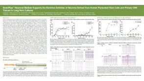 科学海报BrainPhys™ Neuronal Medium Supports the Electrical Activities of Neurons Derived from Human Pluripotent Stem Cells and Primary CNS Tissues in Long-Term Cultures
 沪公网安备31010102008431号