Lee S-HH et al. (JUN 2000)
Nature biotechnology 18 6 675--9
Efficient generation of midbrain and hindbrain neurons from mouse embryonic stem cells.
Embryonic stem (ES) cells are clonal cell lines derived from the inner cell mass of the developing blastocyst that can proliferate extensively in vitro and are capable of adopting all the cell fates in a developing embryo. Clinical interest in the use of ES cells has been stimulated by studies showing that isolated human cells with ES properties from the inner cell mass or developing germ cells can provide a source of somatic precursors. Previous studies have defined in vitro conditions for promoting the development of specific somatic fates,specifically,hematopoietic,mesodermal,and neurectodermal. In this study,we present a method for obtaining dopaminergic (DA) and serotonergic neurons in high yield from mouse ES cells in vitro. Furthermore,we demonstrate that the ES cells can be obtained in unlimited numbers and that these neuron types are generated efficiently. We generated CNS progenitor populations from ES cells,expanded these cells and promoted their differentiation into dopaminergic and serotonergic neurons in the presence of mitogen and specific signaling molecules. The differentiation and maturation of neuronal cells was completed after mitogen withdrawal from the growth medium. This experimental system provides a powerful tool for analyzing the molecular mechanisms controlling the functions of these neurons in vitro and in vivo,and potentially for understanding and treating neurodegenerative and psychiatric diseases.
View Publication
产品号#:
06902
06952
07152
07157
00321
00322
00323
00324
00325
产品名:
N2 添加物-A
Jin HK et al. (MAY 2002)
The Journal of clinical investigation 109 9 1183--91
Intracerebral transplantation of mesenchymal stem cells into acid sphingomyelinase-deficient mice delays the onset of neurological abnormalities and extends their life span.
Types A and B Niemann-Pick disease (NPD) are lysosomal storage disorders resulting from loss of acid sphingomyelinase (ASM) activity. We have used a knockout mouse model of NPD (ASMKO mice) to evaluate the effects of direct intracerebral transplantation of bone marrow-derived mesenchymal stem cells (MSCs) on the progression of neurological disease in this disorder. MSCs were transduced with a retroviral vector to overexpress ASM and were injected into the hippocampus and cerebellum of 3-week-old ASMKO pups. Transplanted cells migrated away from the injection sites and survived at least 6 months after transplantation. Seven of 8 treated mice,but none of the untreated controls,survived for textgreater or = 7 months after transplant. Survival times were greater in sex-matched than in sex-mismatched transplants. Transplantation significantly delayed the Purkinje cell loss that is characteristic of NPD,although the protective effect declined with distance from the injection site. Overall ASM activity in brain homogenates was low,but surviving Purkinje cells contained the retrovirally expressed human enzyme,and transplanted animals showed a reduction in cerebral sphingomyelin. These results reveal the potential of treating neurodegenerative lysosomal storage disorders by intracerebral transplantation of bone marrow-derived MSCs.
View Publication
产品号#:
05350
产品名:
Ohtsuka T et al. (JAN 2006)
Molecular and cellular neurosciences 31 1 109--22
Visualization of embryonic neural stem cells using Hes promoters in transgenic mice.
In the central nervous system,neural stem cells proliferate in the ventricular zone (VZ) and sequentially give rise to both neurons and glial cells in a temporally and spatially regulated manner,suggesting that stem cells may differ from one another in different brain regions and at different developmental stages. For the purpose of marking and purifying neural stem cells to ascertain whether such differences exist,we generated transgenic mice using promoters from Hes genes (pHes1 or pHes5) to drive expression of destabilized enhanced green fluorescent protein. In the developing brains of these transgenic mice,GFP expression was restricted to undifferentiated cells in the VZ,which could asymmetrically produce a Numb-positive neuronal daughter and a GFP-positive progenitor cell in clonal culture,indicating that they retain the capacity to self-renew. Our results suggest that pHes-EGFP transgenic mice can be used to explore similarities and differences among neural stem cells during development.
View Publication
产品号#:
05700
05701
05702
产品名:
NeuroCult™ 基础培养基(小鼠和大鼠)
NeuroCult™ 扩增添加物(小鼠和大鼠)
NeuroCult™扩增试剂盒(小鼠和大鼠)
Kucia M et al. (JAN 2006)
Leukemia 20 1 18--28
Cells enriched in markers of neural tissue-committed stem cells reside in the bone marrow and are mobilized into the peripheral blood following stroke.
The concept that bone marrow (BM)-derived cells participate in neural regeneration remains highly controversial and the identity of the specific cell type(s) involved remains unknown. We recently reported that the BM contains a highly mobile population of CXCR4+ cells that express mRNA for various markers of early tissue-committed stem cells (TCSCs),including neural TCSCs. Here,we report that these cells not only express neural lineage markers (beta-III-tubulin,Nestin,NeuN,and GFAP),but more importantly form neurospheres in vitro. These neural TCSCs are present in significant amounts in BM harvested from young mice but their abundance and responsiveness to gradients of motomorphogens,such as SDF-1,HGF,and LIF,decreases with age. FACS analysis,combined with analysis of neural markers at the mRNA and protein levels,revealed that these cells reside in the nonhematopoietic CXCR4+/Sca-1+/lin-/CD45 BM mononuclear cell fraction. Neural TCSCs are mobilized into the peripheral-blood following stroke and chemoattracted to the damaged neural tissue in an SDF-1-CXCR4-,HGF-c-Met-,and LIF-LIF-R-dependent manner. Based on these data,we hypothesize that the postnatal BM harbors a nonhematopoietic population of cells that express markers of neural TCSCs that may account for the beneficial effects of BM-derived cells in neural regeneration.
View Publication
产品号#:
05700
05701
05702
05703
05704
产品名:
NeuroCult™ 基础培养基(小鼠和大鼠)
NeuroCult™ 扩增添加物(小鼠和大鼠)
NeuroCult™扩增试剂盒(小鼠和大鼠)
NeuroCult™ 分化添加物(小鼠和大鼠)
NeuroCult™ 分化试剂盒(小鼠和大鼠)
Kucia M et al. (JUL 2005)
Leukemia 19 7 1118--27
Bone marrow as a home of heterogenous populations of nonhematopoietic stem cells.
Evidence is presented that bone marrow (BM) in addition to CD45(positive) hematopoietic stem cells contains a rare population of heterogenous CD45(negative) nonhematopoietic tissue committed stem cells (TCSC). These nonhematopoietic TCSC (i) are enriched in population of CXCR4(+) CD34(+) AC133(+) lin(-) CD45(-) and CXCR4(+) Sca-1(+) lin(-) CD45(-) in humans and mice,respectively,(ii) display several markers of pluripotent stem cells (PSC) and (iii) as we envision are deposited in BM early in development. Thus,since BM contains versatile nonhematopoietic stem cells,previous studies on plasticity trans-dedifferentiation of BM-derived hematopoietic stem cells (HSC) that did not include proper controls to exclude this possibility could lead to wrong interpretations. Therefore,in this spotlight review we present this alternative explanation of 'plasticity' of BM-derived stem cells based on the assumption that BM stem cells are heterogenous. We also discuss a potential relationship of TCSC/PSC identified by us with other BM-derived CD45(negative) nonhematopoietic stem cells that were recently identified by other investigators (eg MSC,MAPC,USSC and MIAMI cells). Finally,we discuss perspectives and pitfalls in potential application of these cells in regenerative medicine.
View Publication
产品号#:
05700
05701
05702
05703
05704
产品名:
NeuroCult™ 基础培养基(小鼠和大鼠)
NeuroCult™ 扩增添加物(小鼠和大鼠)
NeuroCult™扩增试剂盒(小鼠和大鼠)
NeuroCult™ 分化添加物(小鼠和大鼠)
NeuroCult™ 分化试剂盒(小鼠和大鼠)
Coksaygan T et al. (FEB 2006)
Experimental neurology 197 2 475--85
Neurogenesis in Talpha-1 tubulin transgenic mice during development and after injury.
Talpha-1 tubulin promoter-driven EYFP expression is seen in murine neurons born as early as E9.5. Double labeling with markers for stem cells (Sox 1,Sox 2,nestin),glial progenitors (S100beta,NG2,Olig2),and neuronal progenitors (doublecortin,betaIII-tubulin,PSA-NCAM) show that Talpha-1 tubulin expression is limited to early born neurons. BrdU uptake and double labeling with neuronal progenitor markers in vivo and in vitro show that EYFP-expressing cells are postmitotic and Talpha-1 tubulin EYFP precedes the expression of MAP-2 and NeuN,and follows the expression of PSA-NCAM,doublecortin (Dcx),and betaIII-tubulin. Talpha-1 tubulin promoter-driven EYFP expression is transient and disappears in most neurons by P0. Persistent EYFP expression is mainly limited to scattered cells in the subventricular zone (SVZ),rostral migratory stream,and hippocampus. However,there are some areas that continue to express Talpha-1 tubulin in the adult without apparent neurogenesis. The number of EYFP-expressing cells declines with age indicating that Talpha-1 tubulin accurately identifies early born postmitotic neurons throughout development but less clearly in the adult. Assessment of neurogenesis after stab wound injuries in the cortex,cerebellum and spinal cord of adult animals shows no neurogenesis in most areas with an increase in BrdU incorporation in glial and other non neuronal populations. An up-regulation of Talpha-1 tubulin can be seen in certain areas unaccompanied by new neurogenesis. Our results suggest that even if stem cells proliferate their ability to generate neurons is limited and caution is warranted in attributing increased BrdU incorporation to stem cells or cells fated to be neurons even in neurogenic areas.
View Publication
产品号#:
05700
05701
05702
产品名:
NeuroCult™ 基础培养基(小鼠和大鼠)
NeuroCult™ 扩增添加物(小鼠和大鼠)
NeuroCult™扩增试剂盒(小鼠和大鼠)
Kim S-J et al. (MAY 2006)
Human molecular genetics 15 10 1580--6
Palmitoyl-protein thioesterase-1 deficiency leads to the activation of caspase-9 and contributes to rapid neurodegeneration in INCL.
The infantile neuronal ceroid lipofuscinosis (INCL),a rare (one in 100 000 births) but one of the most lethal inherited neurodegenerative storage disorders of childhood,is caused by inactivating mutations in the palmitoyl-protein thioesterase-1 (PPT1) gene. PPT1 cleaves thioester linkages in s-acylated (palmitoylated) proteins and facilitates their degradation and/or recycling. Thus,PPT1-deficiency leads to an abnormal intracellular accumulation of s-acylated proteins causing INCL pathogenesis. Although neuronal apoptosis is the suggested cause of neurodegeneration in this disease,the molecular mechanism(s) remains poorly understood. We recently reported that one of the major pathways of neuronal apoptosis in PPT1-knockout (PPT1-KO) mice that mimic INCL,is mediated by endoplasmic reticulum (ER) stress-induced caspase-12 activation. ER stress also increases the production of reactive oxygen species (ROS),disrupts Ca(2+) homeostasis and increases the potential for destabilizing mitochondrial membrane. Mitochondrial membrane destabilization activates caspase-9 present in this organelle,and can mediate apoptosis. We report here that the levels of superoxide dismutase (SOD),most likely induced by ROS,in human INCL as well as PPT1-KO mouse brain tissues are markedly elevated. Moreover,we demonstrate that activated caspase-3 and cleaved-PARP,indicative of apoptosis,are also increased in these tissues. Using cultured neurospheres from PPT1-KO and wild-type mouse fetuses,we further demonstrate that the levels of ROS,SOD-2,cleaved-caspase-9,activated caspase-3 and cleaved-PARP are elevated. We propose that: (i) ER stress due to PPT1-deficiency increases ROS and disrupts calcium homeostasis activating caspase-9 and (ii) caspase-9 activation mediates caspase-3 activation and apoptosis contributing to rapid neurodegeneration in INCL.
View Publication
产品号#:
05700
05701
05702
产品名:
NeuroCult™ 基础培养基(小鼠和大鼠)
NeuroCult™ 扩增添加物(小鼠和大鼠)
NeuroCult™扩增试剂盒(小鼠和大鼠)
Mizutani E et al. (DEC 2006)
Reproduction (Cambridge,England) 132 6 849--57
Developmental ability of cloned embryos from neural stem cells.
The success rate is generally higher when cloning mice from embryonic stem (ES) cell nuclei than from somatic cell nuclei,suggesting that the embryonic nature or the undifferentiated state of the donor cell increases cloning efficiency. We assessed the developmental ability of cloned embryos derived from cultured neural stem cell (NSC) nuclei and compared the success rate with that of embryos cloned from other donor cells such as differentiated NSCs,cumulus cells,Sertoli cells and ES cells in the mouse. The transfer of two-cell cloned embryos derived from cultured NSC nuclei into surrogate mothers produced five live cloned mice. However,the success rate (0.5%) was higher in embryos cloned from cultured NSC nuclei than from differentiated NSCs (0%),but lower than that obtained by cloning mice from other cell nuclei (2.2-3.5%). Although the in vitro developmental potential to the two-cell stage of the cloned embryos derived from NSC nuclei (73%) was similar to that of the cloned embryos derived from other somatic cell nuclei (e.g.,85% in Sertoli cells and 75% in cumulus cells),the developmental rate to the morula-blastocyst stage was only 7%. This rate is remarkably lower than that produced from other somatic cells (e.g.,50% in Sertoli cells and 54% in cumulus cells). These results indicate that the undifferentiated state of neural cells does not enhance the cloning efficiency in mice and that the arrest point for in vitro development of cloned embryos depends on the donor cell type.
View Publication

 EasySep™小鼠TIL(CD45)正选试剂盒


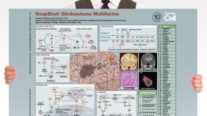 挂图SnapShot: Glioblastoma Multiforme Overview of the key concepts and mechanisms in glioblastoma multiforme biology

 沪公网安备31010102008431号