
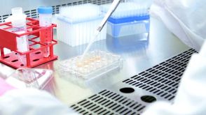
 EasySep™小鼠TIL(CD45)正选试剂盒


技术资料
-
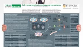 挂图Cell-Reprogramming Technology and Neuroscience Details on human iPSC-derived models of neuropsychiatric and neurodegenerative disorders
 -
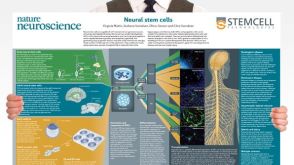 挂图Neural Stem Cells Overview of the types of NSCs and their potential use as therapeutic agents for disease
 -

-
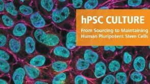 产品手册hPSC 培养 人多能干细胞的来源和维持培养
品牌:
AggreWell,BrainPhys,CryoStor,EasySep,PBS-MINI,ReLeSR,STEMdiff,TeSR,mFreSR
-


 沪公网安备31010102008431号