
 EasySep™小鼠TIL(CD45)正选试剂盒


技术资料
-
-
-
-
-
-
 27:19
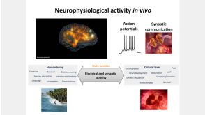
线上讲座BrainPhys™ Medium Supports the Physiological Activity of Neuronal Tissue in vitro发布日期: 07/22/2016
27:19
线上讲座BrainPhys™ Medium Supports the Physiological Activity of Neuronal Tissue in vitro发布日期: 07/22/2016

 沪公网安备31010102008431号