Y. Kim et al. (May 2020)
FASEB Journal 34 6965-6983
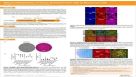
Microtubule-associated protein 2 mediates induction of long-term potentiation in hippocampal neurons
Microtubule-associated protein (MAP) 2 has been perceived as a static cytoskeletal protein enriched in neuronal dendritic shafts. Emerging evidence indicates dynamic functions for various MAPs in activity-dependent synaptic plasticity. However,it is unclear how MAP2 is associated with synaptic plasticity mechanisms. Here,we demonstrate that specific silencing of high-molecular-weight MAP2 in vivo abolished induction of long-term potentiation (LTP) in the Schaffer collateral pathway of CA1 pyramidal neurons and in vitro blocked LTP-induced surface delivery of AMPA receptors and spine enlargement. In mature hippocampal neurons,we observed rapid translocation of a subpopulation of MAP2,present in dendritic shafts,to spines following LTP stimulation. Time-lapse confocal imaging showed that spine translocation of MAP2 was coupled with LTP-induced spine enlargement. Consistently,immunogold electron microscopy revealed that LTP stimulation of the Schaffer collateral pathway promoted MAP2 labeling in spine heads of CA1 neurons. This translocation depended on NMDA receptor activation and Ras-MAPK signaling. Furthermore,LTP stimulation led to an increase in surface-expressed AMPA receptors specifically in the neurons with MAP2 spine translocation. Altogether,this study indicates a novel role for MAP2 in LTP mechanisms and suggests that MAP2 participates in activity-dependent synaptic plasticity in mature hippocampal networks.
View Publication
M. van den Hurk et al. ( 2018)
Frontiers in Molecular Neuroscience
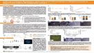
Patch-Seq Protocol to Analyze the Electrophysiology, Morphology and Transcriptome of Whole Single Neurons Derived From Human Pluripotent Stem Cells
The human brain is composed of a complex assembly of about 171 billion heterogeneous cellular units (86 billion neurons and 85 billion non-neuronal glia cells). A comprehensive description of brain cells is necessary to understand the nervous system in health and disease. Recently,advances in genomics have permitted the accurate analysis of the full transcriptome of single cells (scRNA-seq). We have built upon such technical progress to combine scRNA-seq with patch-clamping electrophysiological recording and morphological analysis of single human neurons in vitro. This new powerful method,referred to as Patch-seq,enables a thorough,multimodal profiling of neurons and permits us to expose the links between functional properties,morphology,and gene expression. Here,we present a detailed Patch-seq protocol for isolating single neurons from in vitro neuronal cultures. We have validated the Patch-seq whole-transcriptome profiling method with human neurons generated from embryonic and induced pluripotent stem cells (ESCs/iPSCs) derived from healthy subjects,but the procedure may be applied to any kind of cell type in vitro. Patch-seq may be used on neurons in vitro to profile cell types and states in depth to unravel the human molecular basis of neuronal diversity and investigate the cellular mechanisms underlying brain disorders.
View Publication

 EasySep™小鼠TIL(CD45)正选试剂盒


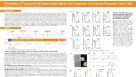
 科学海报Efficient Generation of Human Pluripotent Stem Cells to Neural Crest Cells
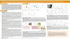


 沪公网安备31010102008431号