
 EasySep™小鼠TIL(CD45)正选试剂盒


技术资料
-

-

-

-
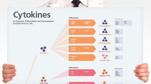 挂图Human Immune Cytokines Infographic of key cytokines for expansion, differentiation and characterization of major immune cell types
 -

-
 科学海报Immunomagnetic Purification of Human Central and Effector Memory T Cell Subsets in 45 Minutes
Conference:
KEYSTONE 2018


 沪公网安备31010102008431号