Szewczyk K et al. (JUN 2016)
Human immunology 77 6 449--55
Flow cytometry crossmatch reactivity with pronase-treated T cells induced by non-HLA autoantibodies in human immunodeficiency virus-infected patients.
Pronase treatment is used in the flow cytometry crossmatch (FCXM) to prevent nonspecific antibody binding on B cells. However,we have observed unexpected positive results with pronase-treated T cells in human immunodeficiency virus (HIV)-infected patients. In this study,25 HIV-infected patients without HLA antibodies were tested with pronase-treated and nontreated cells. HIV-positive sera were pretreated with reducing agents and preabsorbed with pronase-treated and nontreated T or B cells before crossmatching. All patients displayed FCXM reactivity with pronase-treated T cells but not with nontreated T cells. None of the patients exhibited FCXM reactivity with pronase-treated and nontreated B cells. These patients displayed FCXM reactivity with pronase-treated CD4+ and CD8+ T cells but not with their nontreated counterparts. Preabsorption with pronase-treated T cells reduced the T cell FCXM reactivity. Preabsorption with pronase-treated B cells or nontreated T and B cells did not have any effect on the T cell FCXM reactivity. Pretreatment with reducing agents did not affect the T cell FCXM reactivity. 15 of 21 HIV-infected kidney allograft recipients with pronase-treated T cell FCXM reactivity display long-term graft survival (1193±631days). These data indicate that HIV-infected patients have nondeleterious autoantibodies recognizing cryptic epitopes exposed by pronase on T cells.
View Publication
Bosma M et al. (APR 2016)
Nature Communications 7 11314
FNDC4 acts as an anti-inflammatory factor on macrophages and improves colitis in mice.
FNDC4 is a secreted factor sharing high homology with the exercise-associated myokine irisin (FNDC5). Here we report that Fndc4 is robustly upregulated in several mouse models of inflammation as well as in human inflammatory conditions. Specifically,FNDC4 levels are increased locally at inflamed sites of the intestine of inflammatory bowel disease patients. Interestingly,administration of recombinant FNDC4 in the mouse model of induced colitis markedly reduces disease severity compared with mice injected with a control protein. Conversely,mice lacking Fndc4 develop more severe colitis. Analysis of binding of FNDC4 to different immune cell types reveals strong and specific binding to macrophages and monocytes. FNDC4 treatment of bone marrow-derived macrophages in vitro results in reduced phagocytosis,increased cell survival and reduced proinflammatory chemokine expression. Hence,treatment with FNDC4 results in a state of dampened macrophage activity,while enhancing their survival. Thus,we have characterized FNDC4 as a factor with direct therapeutic potential in inflammatory bowel disease and possibly other inflammatory diseases.
View Publication
Balakrishnan K et al. (OCT 2006)
Blood 108 7 2392--8
Forodesine, an inhibitor of purine nucleoside phosphorylase, induces apoptosis in chronic lymphocytic leukemia cells.
Purine nucleoside phosphorylase (PNP) deficiency in humans results in T lymphocytopenia. Forodesine,a potent inhibitor of PNP,was designed based on the transition-state structure stabilized by the enzyme. Previous studies established that forodesine in the presence of deoxyguanosine (dGuo) inhibits the proliferation of T lymphocytes. A phase 1 clinical trial of forodesine in T-cell malignancies demonstrated significant antileukemic activity with an increase in intracellular dGuo triphosphate (dGTP). High accumulation of dGTP in T cells may be dependent on the levels of deoxynucleoside kinases. Because B-cell chronic lymphocytic leukemia (B-CLL) cells have high activity of deoxycytidine kinase (dCK),we hypothesized that these lymphocytes would respond to forodesine. This postulate was tested in primary lymphocytes during in vitro investigations. Lymphocytes from 12 patients with CLL were incubated with forodesine and dGuo. These CLL cells showed a wide variation in the accumulation of intracellular dGTP without any effect on other deoxynucleotides. This was associated with DNA damage-induced p53 stabilization,phosphorylation of p53 at Ser15,and activation of p21. The dGTP accumulation was related to induction of apoptosis measured by caspase activation,changes in mitochondrial membrane potential,and PARP cleavage. Based on these data,a phase 2 clinical trial of forodesine has been initiated for CLL patients.
View Publication
Yu J-J et al. (FEB 2010)
Clinical and vaccine immunology : CVI 17 2 215--22
Francisella tularensis T-cell antigen identification using humanized HLA-DR4 transgenic mice.
There is no licensed vaccine against the intracellular pathogen Francisella tularensis. The use of conventional mouse strains to screen protective vaccine antigens may be problematic,given the differences in the major histocompatibility complex (MHC) binding properties between murine and human antigen-presenting cells. We used engineered humanized mice that lack endogenous MHC class II alleles but that express a human HLA allele (HLA-DR4 transgenic [tg] mice) to identify potential subunit vaccine candidates. Specifically,we applied a biochemical and immunological screening approach with bioinformatics to select putative F. tularensis subsp. novicida T-cell-reactive antigens using humanized HLA-DR4 tg mice. Cell wall- and membrane-associated proteins were extracted with Triton X-114 detergent and were separated by fractionation with a Rotofor apparatus and whole-gel elution. A series of proteins were identified from fractions that stimulated antigen-specific gamma interferon (IFN-gamma) production,and these were further downselected by the use of bioinformatics and HLA-DR4 binding algorithms. We further examined the validity of this combinatorial approach with one of the identified proteins,a 19-kDa Francisella tularensis outer membrane protein (designated Francisella outer membrane protein B [FopB]; FTN0119). FopB was shown to be a T-cell antigen by a specific IFN-gamma recall assay with purified CD4(+) T cells from F. tularensis subsp. novicida DeltaiglC-primed HLA-DR4 tg mice and cells of a human B-cell line expressing HLA-DR4 (DRB1*0401) functioning as antigen-presenting cells. Intranasal immunization of HLA-DR4 tg mice with the single antigen FopB conferred significant protection against lethal pulmonary challenge with an F. tularensis subsp. holarctica live vaccine strain. These results demonstrate the value of combining functional biochemical and immunological screening with humanized HLA-DR4 tg mice to map HLA-DR4-restricted Francisella CD4(+) T-cell epitopes.
View Publication
From blood monocytes to adipose tissue-resident macrophages: induction of diapedesis by human mature adipocytes.
Obesity has been suggested to be a low-grade systemic inflammatory state,therefore we studied the interaction between human adipocytes and monocytes via adipose tissue (AT)-derived capillary endothelium. Cells composing the stroma-vascular fraction (SVF) of human ATs were characterized by fluorescence-activated cell sorter (FACS) analysis and two cell subsets (resident macrophages and endothelial cells [ECs]) were isolated using antibody-coupled microbeads. Media conditioned by mature adipocytes maintained in fibrin gels were applied to AT-derived ECs. Thereafter,the expression of endothelial adhesion molecules was analyzed as well as the adhesion and transmigration of human monocytes. FACS analysis showed that 11% of the SVF is composed of CD14(+)/CD31(+) cells,characterized as resident macrophages. A positive correlation was found between the BMI and the percentage of resident macrophages,suggesting that fat tissue growth is associated with a recruitment of blood monocytes. Incubation of AT-derived ECs with adipocyte-conditioned medium resulted in the upregulation of EC adhesion molecules and the increased chemotaxis of blood monocytes,an effect mimicked by recombinant human leptin. These results indicate that adipokines,such as leptin,activate ECs,leading to an enhanced diapedesis of blood monocytes,and suggesting that fat mass growth might be linked to inflammatory processes.
View Publication
Castriconi R et al. (JUN 2007)
Blood 109 11 4873--81
Functional characterization of natural killer cells in type I leukocyte adhesion deficiency.
In this study,we analyzed IL-2-activated polyclonal natural killer (NK) cells derived from 2 patients affected by leukocyte adhesion deficiency type I (LAD1),an immunodeficiency characterized by mutations of the gene coding for CD18,the beta subunit shared by major leukocyte integrins. We show that LAD1 NK cells express normal levels of various triggering NK receptors (and coreceptors) and that mAb-mediated engagement of these receptors results in the enhancement of both NK cytolytic activity and cytokine production. Moreover,these activating NK receptors were capable of recognizing their specific ligands on target cells. Thus,LAD1 NK cells,similarly to normal NK cells,were capable of killing most human tumor cells analyzed and produced high amounts of IFN-gamma when cocultured in presence of target cells. Murine target cells represented a common exception,as they were poorly susceptible to LAD1 NK cells. Finally,LAD1 NK cells could efficiently kill or induce maturation of monocyte-derived immature dendritic cells (DCs). Altogether our present study indicates that in LAD1 patients,3 important functions of NK cells (eg,cytotoxicity,IFN-gamma production,and DC editing) are only marginally affected and provides new insight on the cooperation between activating receptors and LFA-1 in the induction of NK cell activation and function.
View Publication
Chen H et al. (DEC 2015)
Biological research 48 1 59
Functional disruption of human leukocyte antigen II in human embryonic stem cell.
BACKGROUND Theoretically human embryonic stem cells (hESCs) have the capacity to self-renew and differentiate into all human cell types. Therefore,the greatest promise of hESCs-based therapy is to replace the damaged tissues of patients suffering from traumatic or degenerative diseases by the exact same type of cells derived from hESCs. Allograft immune rejection is one of the obstacles for hESCs-based clinical applications. Human leukocyte antigen (HLA) II leads to CD4(+) T cells-mediated allograft rejection. Hence,we focus on optimizing hESCs for clinic application through gene modification. RESULTS Transcription activator-like effector nucleases (TALENs) were used to target MHC class II transactivator (CIITA) in hESCs efficiently. CIITA (-/-) hESCs did not show any difference in the differentiation potential and self-renewal capacity. Dendritic cells (DCs) derived from CIITA (-/-) hESCs expressed CD83 and CD86 but without the constitutive HLA II. Fibroblasts derived from CIITA (-/-) hESCs were powerless in IFN-$\$ expression of HLA II. CONCLUSION We generated HLA II defected hESCs via deleting CIITA,a master regulator of constitutive and IFN-$\$ expression of HLA II genes. CIITA (-/-) hESCs can differentiate into tissue cells with non-HLA II expression. It's promising that CIITA (-/-) hESCs-derived cells could be used in cell therapy (e.g.,T cells and DCs) and escape the attack of receptors' CD4(+) T cells,which are the main effector cells of cellular immunity in allograft.
View Publication
Milush JM et al. (NOV 2009)
Blood 114 23 4823--31
Functionally distinct subsets of human NK cells and monocyte/DC-like cells identified by coexpression of CD56, CD7, and CD4.
The lack of natural killer (NK) cell-specific markers,as well as the overlap among several common surface antigens and functional properties,has obscured the delineation between NK cells and dendritic cells. Here,novel subsets of peripheral blood CD3/14/19(neg) NK cells and monocyte/dendritic cell (DC)-like cells were identified on the basis of CD7 and CD4 expression. Coexpression of CD7 and CD56 differentiates NK cells from CD56+ monocyte/DC-like cells,which lack CD7. In contrast to CD7+CD56+ NK cells,CD7(neg)CD56+ cells lack expression of NK cell-associated markers,but share commonalities in their expression of various monocyte/DC-associated markers. Using CD7,we observed approximately 60% of CD4+CD56+ cells were CD7(neg) cells,indicating the actual frequency of activated CD4+ NK cells is much lower in the blood than previously recognized. Functionally,only CD7+ NK cells secrete gamma interferon (IFNgamma) and degranulate after interleukin-12 (IL-12) plus IL-18 or K562 target cell stimulation. Furthermore,using CD7 to separate CD56+ NK cells and CD56+ myeloid cells,we demonstrate that unlike resting CD7+CD56+ NK cells,the CD7(neg)CD56+ myeloid cells stimulate a potent allogeneic response. Our data indicate that CD7 and CD56 coexpression discriminates NK cells from CD7(neg)CD56+ monocyte/DC-like cells,thereby improving our ability to study the intricacies of NK-cell subset phenotypes and functions in vivo.
View Publication
Y. Kuwano et al. (MAY 2016)
Journal of Immunology 196 9 3828--33
G$\alpha$i2 and G$\alpha$i3 Differentially Regulate Arrest from Flow and Chemotaxis in Mouse Neutrophils.
Leukocyte recruitment to inflammation sites progresses in a multistep cascade. Chemokines regulate multiple steps of the cascade,including arrest,transmigration,and chemotaxis. The most important chemokine receptor in mouse neutrophils is CXCR2,which couples through G$\alpha$i2- and G$\alpha$i3-containing heterotrimeric G proteins. Neutrophils arrest in response to CXCR2 stimulation. This is defective in G$\alpha$i2-deficient neutrophils. In this study,we show that G$\alpha$i3-deficient neutrophils showed reduced transmigration but normal arrest in mice. We also tested G$\alpha$i2- or G$\alpha$i3-deficient neutrophils in a CXCL1 gradient generated by a microfluidic device. G$\alpha$i3-,but not G$\alpha$i2-,deficient neutrophils showed significantly reduced migration and directionality. This was confirmed in a model of sterile inflammation in vivo. G$\alpha$i2-,but not G$\alpha$i3-,deficient neutrophils showed decreased Ca(2+) flux in response to CXCR2 stimulation. Conversely,G$\alpha$i3-,but not G$\alpha$i2-,deficient neutrophils exhibited reduced AKT phosphorylation upon CXCR2 stimulation. We conclude that G$\alpha$i2 controls arrest and G$\alpha$i3 controls transmigration and chemotaxis in response to chemokine stimulation of neutrophils.
View Publication

 EasySep™小鼠TIL(CD45)正选试剂盒


 挂图Frequencies and Percentages of Mouse Immune Cell Types List of the frequencies of over 25 immune cell types in C57BL/6 mice
 挂图Frequencies of Immune Cells in Rat Tissue Lists the estimated frequencies of more than 15 immune cell types in Sprague Dawley rats
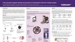 科学海报Fully Automated Magnetic Labeling and Separation of Hematopoietic Cells from Multiple Samples
 沪公网安备31010102008431号