Geldanamycin and herbimycin A induce apoptotic killing of B chronic lymphocytic leukemia cells and augment the cells' sensitivity to cytotoxic drugs.
We studied the actions of geldanamycin (GA) and herbimycin A (HMA),inhibitors of the chaperone proteins Hsp90 and GRP94,on B chronic lymphocytic leukemia (CLL) cells in vitro. Both drugs induced apoptosis of the majority of CLL isolates studied. Whereas exposure to 4-hour pulses of 30 to 100 nM GA killed normal B lymphocytes and CLL cells with similar dose responses,T lymphocytes from healthy donors as well as those present in the CLL isolates were relatively resistant. GA,but not HMA,showed a modest cytoprotective effect toward CD34+ hematopoietic progenitors from normal bone marrow. The ability of bone marrow progenitors to form hematopoietic colonies was unaffected by pulse exposures to GA. Both GA and HMA synergized with chlorambucil and fludarabine in killing a subset of CLL isolates. GA- and HMA-induced apoptosis was preceded by the up-regulation of the stress-responsive chaperones Hsp70 and BiP. Both ansamycins also resulted in down-regulation of Akt protein kinase,a modulator of cell survival. The relative resistance of T lymphocytes and of CD34+ bone marrow progenitors to GA coupled with its ability to induce apoptosis following brief exposures and to synergize with cytotoxic drugs warrant further investigation of ansamycins as potential therapeutic agents in CLL.
View Publication
产品号#:
04434
04444
产品名:
MethoCult™ H4434 Classic
MethoCult™ H4434 Classic
Coffman KT et al. (NOV 2003)
Cancer Research 63 22 7907--12
Differential EphA2 epitope display on normal versus malignant cells.
The EphA2 receptor tyrosine kinase is overexpressed in many different types of human cancers where it functions as a powerful oncoprotein. Dramatic changes in the subcellular localization and function of EphA2 have also been linked with cancer,and in particular,unstable cancer cell-cell contacts prevent EphA2 from stably binding its ligand on the surface of adjoining cells. This change is important in light of evidence that ligand binding causes EphA2 to transmit signals that negatively regulate tumor cell growth and invasiveness and also induce EphA2 degradation. On the basis of these properties,we have begun to target EphA2 on tumor cells using agonistic antibodies,which mimic the consequences of ligand binding. In our present study,we show that a subset of agonistic EphA2 antibodies selectively bind epitopes on malignant cells,which are not available on nontransformed epithelial cells. We also show that such epitopes arise from differential cell-cell adhesions and that the stable intercellular junctions of nontransformed epithelial cells occlude the binding site for ligand,as well as this subset of EphA2 antibodies. Finally,we demonstrate that antibody targeting of EphA2 decreases tumor cell growth as measured using xenograft tumor models and found that the mechanism of antibody action relates to EphA2 protein degradation in vivo. Taken together,these results suggest new opportunities for therapeutic targeting of the large number of different cancers that express EphA2 in a manner that could minimize potential toxicities to normal cells.
View Publication
产品号#:
03800
03801
03802
03803
03804
03805
03806
产品名:
ClonaCell™-HY杂交瘤试剂盒
ClonaCell™-HY培养基A
ClonaCell™-HY 培养基 B
ClonaCell™-HY 培养基 C
ClonaCell™-HY 培养基 D
ClonaCell™-HY 培养基 E
ClonaCell™-HY PEG
Niedre MJ et al. (NOV 2003)
Cancer research 63 22 7986--94
In vitro tests of the validity of singlet oxygen luminescence measurements as a dose metric in photodynamic therapy.
Singlet oxygen ((1)O(2)) is widely believed to be the major cytotoxic agent involved in photodynamic therapy (PDT). We showed recently that measurement of the weak near infrared luminescence of (1)O(2) is possible in cells in vitro and tissues in vivo. Here,we investigated the relationship between the integrated luminescence signal and the in vitro PDT response of AML5 leukemia cells sensitized with aminolevulinic acid-induced protoporphyrin IX (PpIX). Sensitized cell suspensions were irradiated with pulsed 523 nm laser light at average fluence rates of 10,25,or 50 mWcm(-2) and,(1)O(2) luminescence measurements were made throughout the treatment. Cell survival was measured with either propidium iodide-labeled flow cytometry or colony-forming assay. The PpIX concentration in the cells,the photobleaching,and the pO(2) in the cell suspensions were also monitored. There were large variations in cell survival and (1)O(2) generation in different experiments due to different controlled treatment parameters (fluence and fluence rate) and other uncontrolled factors (PpIX synthesis and oxygenation). However,in all of the cases,cell kill correlated strongly with the cumulative (1)O(2) luminescence and allowed direct estimation of the (1)O(2) per cell required to achieve a specific level of cell kill. This study supports the validity and potential utility of (1)O(2) luminescence measurement as a dosimetric tool for PDT,as well as confirming the likely role of (1)O(2) in porphyrin-based PDT.
View Publication
产品号#:
04531
产品名:
MethoCult™ H4531
Hideshima T et al. (DEC 2003)
Cancer research 63 23 8428--36
Antitumor activity of lysophosphatidic acid acyltransferase-beta inhibitors, a novel class of agents, in multiple myeloma.
In this study,we examined the effects of isoform-specific functional inhibitors of lysophosphatidic acid acyltransferase (LPAAT),which converts lysophosphatidic acid to phosphatidic acid,on multiple myeloma (MM) cell growth and survival. The LPAAT-beta inhibitors CT-32176,CT-32458,and CT-32615 induced textgreater95% growth inhibition (P textless 0.01) in MM.1S,U266,and RPMI8226 MM cell lines,as well as MM cells from patients (IC(50),50-200 nM). We further characterized this LPAAT-beta inhibitory effect using CT-32615,the most potent inhibitor of MM cell growth. CT-32615 triggered apoptosis in MM cells via caspase-8,caspase-3,caspase-7,and poly (ADP-ribose) polymerase cleavage. Neither interleukin 6 nor insulin-like growth factor I inhibited CT-32615-induced apoptosis. Dexamethasone and immunomodulatory derivatives of thalidomide (IMiDs),but not proteasome inhibitor PS-341,augmented MM cell apoptosis triggered by LPAAT-beta inhibitors. CT-32615-induced apoptosis was associated with phosphorylation of p53 and c-Jun NH(2)-terminal kinase (JNK); conversely,JNK inhibitor SP600125 and dominant-negative JNK inhibited CT-32615-induced apoptosis. Importantly,CT-32615 inhibited tumor necrosis factor-alpha-triggered nuclear factor-kappaB activation but did not affect either tumor necrosis factor-alpha-induced p38 mitogen-activated protein kinase phosphorylation or interleukin 6-triggered signal transducers and activators of transcription 3 phosphorylation. Finally,although binding of MM cells to bone marrow stromal cells augments MM cell growth and protects against dexamethasone-induced apoptosis,CT-32615 induced apoptosis even of adherent MM cells. Our data therefore demonstrate for the first time that inhibiting LPAAT-beta induces cytotoxicity in MM cells in the bone marrow milieu,providing the framework for clinical trials of these novel agents in MM.
View Publication
产品号#:
15129
15169
产品名:
RosetteSep™人多发性骨髓瘤细胞富集抗体混合物
RosetteSep™人多发性骨髓瘤细胞富集抗体混合物
Li EC and Davis LE (NOV 2003)
Clinical therapeutics 25 11 2669--708
Zoledronic acid: a new parenteral bisphosphonate.
BACKGROUND Inhibition of bone resorption using bisphosphonates is an important step in palliation of complications of advanced cancer,such as hypercalcemia and metastatic bone disease. OBJECTIVE The goal of this article was to describe the pharmacologic properties of zoledronic acid (zoledronate) and discuss findings from preclinical and clinical studies of its use in skeletal disorders. METHODS Relevant English-language literature was identified using the terms zoledronic acid,zoledronate,Zometa,and 118072-93-8 through searches of MEDLINE (1966-June 2003) and International Pharmaceutical Abstracts (1970-June 2003),and abstract proceedings from the American Society of Clinical Oncology (1997-2002). RESULTS Zoledronic acid is a nitrogen-containing bisphosphonate that inhibits bone resorption. It is indicated for the treatment of hypercalcemia of malignancy and for the treatment of patients with multiple myeloma or documented metastasis from solid tumors,in conjunction with standard antineoplastic therapy. The recommended dosage is 4 mg via IV over textgreateror= 15 minutes every 3 or 4 weeks. Compared with pamidronate 90 mg,zoledronic acid 4 and 8 mg provided a higher complete response rate for hypercalcemia of malignancy by day 10 (88.4% and 86.7% vs 69.7%; P = 0.002 and P = 0.015) and longer duration of action (median time to relapse,30 and 40 days vs 17 days; P = 0.001 and P = 0.007). In patients with breast cancer or multiple myeloma,zoledronic acid was as effective as pamidronate in delaying time to a first skeletal-related event (373 days vs 363 days). In patients with hormone-refractory prostate cancer and bone metastases,zoledronic acid 4 mg reduced the proportion of patients who experienced a skeletal-related event (33% vs 44% with placebo; P = 0.021) or a skeletal fracture (13% vs 22% with placebo; P = 0.015). In patients with bone metastases from solid tumors,zoledronic acid delayed the median time to a first skeletal-related event (230 days vs 163 days with placebo; P = 0.023). Common adverse events include fever,nausea,constipation,fatigue,and bone pain. CONCLUSION Zoledronic acid is an effective and generally well-tolerated treatment for hypercalcemia of malignancy and skeletal complications of metastatic bone disease.
View Publication

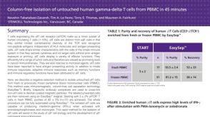
 EasySep™小鼠TIL(CD45)正选试剂盒


 科学海报Mammosphere Culture Supports Short But Not Long-Term Propagation of Human Mammary Epithelial Progenitors
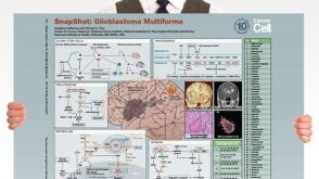 挂图SnapShot: Glioblastoma Multiforme Overview of the key concepts and mechanisms in glioblastoma multiforme biology


 沪公网安备31010102008431号