Nicolini FE et al. (AUG 2002)
Blood 100 4 1257--64
Expression of a human beta-globin transgene in erythroid cells derived from retrovirally transduced transplantable human fetal liver and cord blood cells.
Transfer of therapeutic genes to human hematopoietic stem cells (HSCs) using complex vectors at clinically relevant efficiencies remains a major challenge. Recently we described a stable retroviral vector that sustains long-term expression of green fluorescent protein (GFP) and a human beta-globin gene in the erythroid progeny of transduced murine HSCs. We now report the efficient transduction of primitive human CD34(+) fetal liver or cord blood cells with this vector and expression of the beta-globin transgene in the erythroid progeny of these human cells for at least 2 months. After growth factor prestimulation and then a 2- to 3-day exposure to the virus,35% to 55% GFP(+) progeny were seen in assays of transduced colony-forming cells,primitive erythroid precursors that generate large numbers of glycophorin A(+) cells in 3-week suspension cultures,and 6-week long-term culture-initiating cells. In immunodeficient mice injected with unselected infected cells,5% to 15% of the human cells regenerated in the marrow (including the erythroid cells) were GFP(+) 3 and 6 weeks after transplantation. Importantly,the numbers of GFP(+) human lymphoid and either granulopoietic or erythroid cells in individual mice 6 weeks after transplantation were significantly correlated,indicative of the initial transduction of human multipotent cells with in vivo repopulating activity. Expression of the transduced beta-globin gene in human cells obtained directly from the mice or after their differentiation into erythroid cells in vitro was demonstrated by reverse transcriptase-polymerase chain reaction using specific primers. These experiments represent a significant step toward the realization of a gene therapy approach for human beta-globin gene disorders.
View Publication
产品号#:
04330
产品名:
MethoCult™H4330
Rosé L et al. (JUL 2002)
Experimental hematology 30 7 729--37
In vitro studies of the combination of imatinib mesylate (Gleevec) and arsenic trioxide (Trisenox) in chronic myelogenous leukemia.
OBJECTIVE: The aim of this study was the preclinical evaluation of imatinib mesylate (Gleevec,formerly STI571) in conjunction with arsenic trioxide (As2O3,Trisenox) for the treatment of chronic myelogenous leukemia (CML). MATERIALS AND METHODS: Tetrazolium-based cell line proliferation assays (MTT assays) were performed to determine the cytotoxicity of As2O3 alone and in combination with imatinib. Cell lines tested in this study were Bcr-Abl-expressing cells (K562,MO7p210,32Dp210) and parental cells (MO7e,32D). Isobologram analysis was performed manually and using the median effect method. In vitro cytotoxicity also was determined in colony-forming assays using CML patient cells. Western blot analysis was performed to detect Bcr-Abl protein levels in K562 cells exposed to As2O3 at graded concentrations. Bcr-Abl protein level kinetics were correlated with cell viability (trypan blue count) and activated caspase-3 detected by flow cytometry. RESULTS: We show additive to synergistic cytotoxicity in Bcr-Abl+ cell lines depending on inhibitory concentrations and cell type. Results obtained by colony-forming assays confirmed the findings in cell line proliferation assays. Flow cytometric detection of activated caspase-3 revealed synergistic activity in K562 cells. Treatment of K562 cells with As2O3 alone led to down-regulation of Bcr-Abl protein within 24 hours,even at low doses. The decline of Bcr-Abl preceded activation of caspase-3 and the loss of viable cells. CONCLUSIONS: Favorable cytotoxicity and proapoptotic activity of imatinib in conjunction with As2O3 and specific down-regulation of Bcr-Abl protein levels by As2O3 in K562 cells indicate that As2O3 in combination with imatinib might be useful for circumventing resistance to imatinib monotherapy.
View Publication
产品号#:
04564
04534
04544
产品名:
入门套件MethoCult™H4534经典无EPO
MethoCult™H4534经典无EPO
MethoCult™H4534经典无EPO
Bruserud &O et al. (JUN 2002)
Haematologica 87 6 584--95
Leptin in human acute myelogenous leukemia: studies of in vivo levels and in vitro effects on native functional leukemia blasts.
BACKGROUND AND OBJECTIVES: Leptin receptors can be expressed by acute myelogenous leukemia (AML) cells,but the functional effects of leptin on native AML blasts have not been characterized in detail. We investigated systemic leptin levels in AML patients and in vitro effects of leptin on cultured AML blasts. DESIGN AND METHODS: Serum leptin levels were compared for patients with untreated AML and healthy controls. Native AML blasts were derived from a large group of consecutive patients,and effects of leptin on proliferation (suspension cultures and colony formation),constitutive cytokine secretion,differentiation and apoptosis regulation were assayed in vitro. RESULTS: Systemic leptin levels were decreased in patients with untreated AML,and leptin levels in acute leukemia patients were not altered during severe chemotherapy-induced cytopenia and complicating febrile neutropenia. In vitro studies demonstrated that leptin increased AML blast release of interleukin (IL) 1beta,IL6,tumor necrosis factor (TNF) alpha and granulocyte-macrophage colony-stimulating factor (GM-CSF). This enhancing effect showed no correlation with CD34 expression and was not dependent on the presence of serum,induction of differentiation or alteration of caspase 3 activity with decreased in vitro apoptosis. Leptin also increased spontaneous AML blast proliferation,whereas divergent effects on blast proliferation were observed in the presence of exogenous cytokines. The in vitro effects were usually observed at concentrations exceeding the systemic levels. INTERPRETATION AND CONCLUSIONS: Our results suggest that systemic leptin levels alone do not have a major influence on native AML blasts,but the systemic levels in combination with local leptin release in the bone marrow may affect the functional characteristics of these cells.
View Publication
Selective elimination of leukemic CD34(+) progenitor cells by cytotoxic T lymphocytes specific for WT1.
Hematologic malignancies such as acute and chronic myeloid leukemia are characterized by the malignant transformation of immature CD34(+) progenitor cells. Transformation is associated with elevated expression of the Wilm's tumor gene encoded transcription factor (WT1). Here we demonstrate that WT1 can serve as a target for cytotoxic T lymphocytes (CTL) with exquisite specificity for leukemic progenitor cells. HLA-A0201- restricted CTL specific for WT1 kill leukemia cell lines and inhibit colony formation by transformed CD34(+) progenitor cells isolated from patients with chronic myeloid leukemia (CML),whereas colony formation by normal CD34(+) progenitor cells is unaffected. Thus,the tissue-specific transcription factor WT1 is an ideal target for CTL-mediated purging of leukemic progenitor cells in vitro and for antigen-specific therapy of leukemia and other WT1-expressing malignancies in vivo.
View Publication
产品号#:
04535
04545
产品名:
MethoCult™H4535富集无EPO
MethoCult™ H4535 Enriched,不含EPO
Brandl M et al. (AUG 1999)
Experimental hematology 27 8 1264--70
Bispecific antibody fragments with CD20 X CD28 specificity allow effective autologous and allogeneic T-cell activation against malignant cells in peripheral blood and bone marrow cultures from patients with B-cell lineage leukemia and lymphoma.
Bispecific antibodies directed against tumor-associated target antigens and to surface receptors mediating T-cell activation,such as the TCR/CD3 complex and the costimulatory receptor CD28,are capable of mediating T-cell activation resulting in tumor cell killing. In this study,we used the B-cell-associated antigens CD19 and CD20 as target structures on human leukemic cells. We found that a combination of bispecific antibody fragments (bsFab2) with target x CD3 and target x CD28 specificity induces vigorous autologous T-cell activation and killing of malignant cells in peripheral blood and bone marrow cultures from patients with chronic lymphocytic leukemia and follicular lymphoma. The bsFab2 targeting CD20 were considerably more effective than those binding to CD19. The colony-forming capacity of treated bone marrow was impaired due to large amounts of tumor necrosis factor alpha produced during bsFab2-induced T-cell activation. Neutralizing tumor necrosis factor alpha antibodies were found to reverse this negative effect without affecting T-cell activation and tumor cell killing. CD20 x CD28 bsFab2,when used alone rather than in combination,markedly improved the recognition of leukemic cells by allogeneic T cells. Therefore,these reagents may be capable of enhancing the immunogenicity of leukemic cells in general and,in particular,of increasing the antileukemic activity of allogeneic donor buffy coat cells in relapsed bone marrow transplanted patients.
View Publication

 EasySep™小鼠TIL(CD45)正选试剂盒


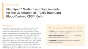 技术公告StemSpan™ Medium and Supplements for the Generation of T Cells from Cord Blood-Derived CD34+ Cells

 沪公网安备31010102008431号