
 EasySep™小鼠TIL(CD45)正选试剂盒


技术资料
-
-
-
 科学海报STEMprep™ Automated Tissue Dissociation System: Maximize Cell Viability and Yield
Conference:
AAI 2026
-
 产品手册Highway1™: Fast, Gentle, and Automated Cell Sorting for Every Lab
品牌:
Highway1
发布日期: 04/14/2026 -
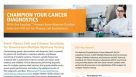
 专家访谈Dr. Theresa Brown Cell Isolation and FISH Testing in a Cytogenetics Lab
研究方向:
癌症研究
发布日期: 01/10/2022


 沪公网安备31010102008431号