D. M. Previte et al. (apr 2019)
Cell reports 27 1 129--141.e4
Lymphocyte Activation Gene-3 Maintains Mitochondrial and Metabolic Quiescence in Naive CD4+ T Cells.
Lymphocyte activation gene-3 (LAG-3) is an inhibitory receptor expressed by CD4+ T cells and tempers their homeostatic expansion. Because CD4+ T cell proliferation is tightly coupled to bioenergetics,we investigate the role of LAG-3 in modulating naive CD4+ T cell metabolism. LAG-3 deficiency enhances the metabolic profile of naive CD4+ T cells by elevating levels of mitochondrial biogenesis. In vivo,LAG-3 blockade partially restores expansion and the metabolic phenotype of wild-type CD4+ T cells to levels of Lag3-/- CD4+ T cells,solidifying that LAG-3 controls these processes. Lag3-/- CD4+ T cells also demonstrate greater signal transducer and activator of transcription 5 (STAT5) activation,enabling resistance to interleukin-7 (IL-7) deprivation. These results implicate this pathway as a target of LAG-3-mediated inhibition. Additionally,enhancement of STAT5 activation,as a result of LAG-3 deficiency,contributes to greater activation potential in these cells. These results identify an additional mode of regulation elicited by LAG-3 in controlling CD4+ T cell responses.
View Publication
文献
S. Omenetti et al. (jun 2019)
Immunity
The Intestine Harbors Functionally Distinct Homeostatic Tissue-Resident and Inflammatory Th17 Cells.
T helper 17 (Th17) cells are pathogenic in many inflammatory diseases,but also support the integrity of the intestinal barrier in a non-inflammatory manner. It is unclear what distinguishes inflammatory Th17 cells elicited by pathogens and tissue-resident homeostatic Th17 cells elicited by commensals. Here,we compared the characteristics of Th17 cells differentiating in response to commensal bacteria (SFB) to those differentiating in response to a pathogen (Citrobacter rodentium). Homeostatic Th17 cells exhibited little plasticity towards expression of inflammatory cytokines,were characterized by a metabolism typical of quiescent or memory T cells,and did not participate in inflammatory processes. In contrast,infection-induced Th17 cells showed extensive plasticity towards pro-inflammatory cytokines,disseminated widely into the periphery,and engaged aerobic glycolysis in addition to oxidative phosphorylation typical for inflammatory effector cells. These findings will help ensure that future therapies directed against inflammatory Th17 cells do not inadvertently damage the resident gut population.
View Publication
文献
B. L. Jamison et al. (jul 2019)
Journal of immunology (Baltimore,Md. : 1950) 203 1 48--57
Nanoparticles Containing an Insulin-ChgA Hybrid Peptide Protect from Transfer of Autoimmune Diabetes by Shifting the Balance between Effector T Cells and Regulatory T Cells.
CD4 T cells play a critical role in promoting the development of autoimmunity in type 1 diabetes. The diabetogenic CD4 T cell clone BDC-2.5,originally isolated from a NOD mouse,has been widely used to study the contribution of autoreactive CD4 T cells and relevant Ags to autoimmune diabetes. Recent work from our laboratory has shown that the Ag for BDC-2.5 T cells is a hybrid insulin peptide (2.5HIP) consisting of an insulin C-peptide fragment fused to a peptide from chromogranin A (ChgA) and that endogenous 2.5HIP-reactive T cells are major contributors to autoimmune pathology in NOD mice. The objective of this study was to determine if poly(lactide-co-glycolide) (PLG) nanoparticles (NPs) loaded with the 2.5HIP Ag (2.5HIP-coupled PLG NPs) can tolerize BDC-2.5 T cells. Infusion of 2.5HIP-coupled PLG NPs was found to prevent diabetes in an adoptive transfer model by impairing the ability of BDC-2.5 T cells to produce proinflammatory cytokines through induction of anergy,leading to an increase in the ratio of Foxp3+ regulatory T cells to IFN-gamma+ effector T cells. To our knowledge,this work is the first to use a hybrid insulin peptide,or any neoepitope,to re-educate diabetogenic T cells and may have significant implications for the development of an Ag-specific therapy for type 1 diabetes patients.
View Publication
文献
L. Hang et al. (apr 2019)
Journal of immunology (Baltimore,Md. : 1950) 202 8 2473--2481
Heligmosomoides polygyrus bakeri Infection Decreases Smad7 Expression in Intestinal CD4+ T Cells, Which Allows TGF-beta to Induce IL-10-Producing Regulatory T Cells That Block Colitis.
Helminthic infections modulate host immunity and may protect their hosts from developing immunological diseases like inflammatory bowel disease. Induction of regulatory T cells (Tregs) may be an important part of this protective process. Heligmosomoides polygyrus bakeri infection also promotes the production of the regulatory cytokines TGF-beta and IL-10 in the gut. In the intestines,TGF-beta helps induce regulatory T cells. This study used Foxp3/IL-10 double reporter mice to investigate the effect of TGF-beta on the differentiation of colon and mesenteric lymph node-derived murine Foxp3- IL-10- CD4+ T cells into their regulatory phenotypes. Foxp3- IL-10- CD4+ T cells from H. polygyrus bakeri-infected mice,as opposed to T cells from uninfected animals,cultured in vitro with TGF-beta and anti-CD3/CD28 mAb differentiated into Foxp3+ and/or IL-10+ T cells. The IL-10-producing T cells nearly all displayed CD25. Smad7 is a natural inhibitor of TGF-beta signaling. In contrast to gut T cells from uninfected mice,Foxp3- IL10- CD4+ T cells from H. polygyrus bakeri-infected mice displayed reduced Smad7 expression and responded to TGF-beta with Smad2/3 phosphorylation. The TGF-beta-induced Tregs that express IL-10 blocked colitis when transferred into the Rag/CD25- CD4+ T cell transfer model of inflammatory bowel disease. TGF-beta had a greatly diminished capacity to induce Tregs in H. polygyrus bakeri-infected transgenic mice with constitutively high T cell-specific Smad7 expression. Thus,infection with H. polygyrus bakeri causes down-modulation in Smad7 expression in intestinal CD4+ T cells,which allows the TGF-beta produced in response to the infection to induce the Tregs that prevent colitis.
View Publication
文献
C. Gu et al. (jul 2019)
Journal of immunology (Baltimore,Md. : 1950) 203 2 389--399
Signaling Cascade through DC-ASGPR Induces Transcriptionally Active CREB for IL-10 Induction and Immune Regulation.
The types and magnitude of Ag-specific immune responses can be determined by the functional plasticity of dendritic cells (DCs). However,how DCs display functional plasticity and control host immune responses have not been fully understood. In this study,we report that ligation of DC-asialoglycoprotein receptor (DC-ASGPR),a C-type lectin receptor (CLR) expressed on human DCs,resulted in rapid activation of Syk,followed by PLCgamma2 and PKCdelta engagements. However,different from other Syk-coupled CLRs,including Dectin-1,signaling cascade through DC-ASGPR did not trigger NF-kappaB activation. Instead,it selectively activated MAPK ERK1/2 and JNK. Rapid and prolonged phosphorylation of ERK1/2 led to sequential activation of p90RSK and CREB,which consequently bound to IL10 promoter and initiated cytokine expression. In addition,DC-ASGPR ligation activated Akt,which differentially regulated the activities of GSK-3alpha/beta and beta-catenin and further contributed to IL-10 expression. Our observations demonstrate that DC-ASGPR induces IL-10 expression via an intrinsic signaling pathway,which provides a molecular explanation for DC-ASGPR-mediated programing of DCs to control host immune responses.
View Publication
文献
E. Giuliani et al. (mar 2019)
Scientific reports 9 1 4373
Hexamethylene bisacetamide impairs NK cell-mediated clearance of acute T lymphoblastic leukemia cells and HIV-1-infected T cells that exit viral latency.
The hexamethylene bisacetamide (HMBA) anticancer drug was dismissed due to limited efficacy in leukemic patients but it may re-enter into the clinics in HIV-1 eradication strategies because of its recently disclosed capacity to reactivate latent virus. Here,we investigated the impact of HMBA on the cytotoxicity of natural killer (NK) cells against acute T lymphoblastic leukemia (T-ALL) cells or HIV-1-infected T cells that exit from latency. We show that in T-ALL cells HMBA upmodulated MICB and ULBP2 ligands for the NKG2D activating receptor. In a primary CD4+ T cell-based latency model,HMBA did not reactivate HIV-1,yet enhanced ULBP2 expression on cells harboring virus reactivated by prostratin (PRO). However,HMBA reduced the expression of NKG2D and its DAP10 adaptor in NK cells,hence impairing NKG2D-mediated cytotoxicity and DAP10-dependent response to IL-15 stimulation. Alongside,HMBA dampened killing of T-ALL targets by IL-15-activated NK cells and impaired NK cell-mediated clearance of PRO-reactivated HIV-1+ cells. Overall,our results demonstrate a dominant detrimental effect of HMBA on the NKG2D pathway that crucially controls NK cell-mediated killing of tumors and virus-infected cells,providing one possible explanation for poor clinical outcome in HMBA-treated cancer patients and raising concerns for future therapeutic application of this drug.
View Publication

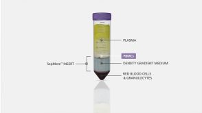
 EasySep™小鼠TIL(CD45)正选试剂盒


 协议Stimulation of Antigen-Specific T Cells Using Peptide Pools
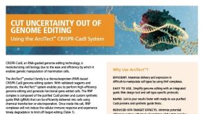 BrochureArciTect™ CRISPR-Cas9 Genome Editing System
 文献
文献


 沪公网安备31010102008431号