Miyoshi H et al. (JAN 1999)
Science (New York,N.Y.) 283 5402 682--6
Transduction of human CD34+ cells that mediate long-term engraftment of NOD/SCID mice by HIV vectors.
Efficient gene transfer into human hematopoietic stem cells (HSCs) is an important goal in the study of the hematopoietic system as well as for gene therapy of hematopoietic disorders. A lentiviral vector based on the human immunodeficiency virus (HIV) was able to transduce human CD34+ cells capable of stable,long-term reconstitution of nonobese diabetic/severe combined immunodeficient (NOD/SCID) mice. High-efficiency transduction occurred in the absence of cytokine stimulation and resulted in transgene expression in multiple lineages of human hematopoietic cells for up to 22 weeks after transplantation.
View Publication
文献
Bystrom J et al. (MAY 2017)
Clinical reviews in allergy & immunology
Response to Treatment with TNFα Inhibitors in Rheumatoid Arthritis Is Associated with High Levels of GM-CSF and GM-CSF(+) T Lymphocytes.
Biologic TNFα inhibitors are a mainstay treatment option for patients with rheumatoid arthritis (RA) refractory to other treatment options. However,many patients either do not respond or relapse after initially responding to these agents. This study was carried out to identify biomarkers that can distinguish responder from non-responder patients before the initiation of treatment. The level of cytokines in plasma and those produced by ex vivo T cells,B cells and monocytes in 97 RA patients treated with biologic TNFα inhibitors was measured before treatment and after 1 and 3 months of treatment by multiplex analyses. The frequency of T cell subsets and intracellular cytokines were determined by flow cytometry. The results reveal that pre-treatment,T cells from patients who went on to respond to treatment with biologic anti-TNFα agents produced significantly more GM-CSF than non-responder patients. Furthermore,immune cells from responder patients produced higher levels of IL-1β,TNFα and IL-6. Cytokine profiling in the blood of patients confirmed the association between high levels of GM-CSF and responsiveness to biologic anti-TNFα agents. Thus,high blood levels of GM-CSF pre-treatment had a positive predictive value of 87.5% (61.6 to 98.5% at 95% CI) in treated RA patients. The study also shows that cells from most anti-TNFα responder patients in the current cohort produced higher levels of GM-CSF and TNFα pre-treatment than non-responder patients. Findings from the current study and our previous observations that non-responsiveness to anti-TNFα is associated with high IL-17 levels suggest that the disease in responder and non-responder RA patients is likely to be driven/sustained by different inflammatory pathways. The use of biomarker signatures of distinct pro-inflammatory pathways could lead to evidence-based prescription of the most appropriate biological therapies for different RA patients.
View Publication
文献
D. Xie et al. (MAY 2017)
Experimental cell research
The effects of activin A on the migration of human breast cancer cells and neutrophils and their migratory interaction.
Activin A belongs to the superfamily of transforming growth factor beta (TGF$\beta$) and is a critical regulatory cytokine in breast cancer and inflammation. However,the role of activin A in migration of breast cancer cells and immune cells was not well characterized. Here,a microfluidic device was used to examine the effect of activin A on the migration of human breast cancer cell line MDA-MB-231 cells and human blood neutrophils as well as their migratory interaction. We found that activin A promoted the basal migration but impaired epidermal growth factor (EGF)-induced migration of breast cancer cells. By contrast,activin A reduced neutrophil chemotaxis and transendothelial migration to N-Formyl-Met-Leu-Phe (fMLP). Finally,activin A promoted neutrophil chemotaxis to the supernatant from breast cancer cell culture. Collectively,our study revealed the different roles of activin A in regulating the migration of breast cancer cells and neutrophils and their migratory interaction. These findings suggested the potential of activin A as a therapeutic target for inflammation and breast cancers.
View Publication
文献
Liu Y-S et al. (MAY 2017)
Oncogene
MiR-181b modulates EGFR-dependent VCAM-1 expression and monocyte adhesion in glioblastoma.
Tumor-associated macrophages (TAMs) originate as circulating monocytes,and are recruited to gliomas,where they facilitate tumor growth and migration. Understanding the interaction between TAM and cancer cells may identify therapeutic targets for glioblastoma multiforme (GBM). Vascular cell adhesion molecule-1 (VCAM-1) is a cytokine-induced adhesion molecule expressed on the surface of cancer cells,which is involved in interactions with immune cells. Analysis of the glioma patient database and tissue immunohistochemistry showed that VCAM-1 expression correlated with the clinico-pathological grade of gliomas. Here,we found that VCAM-1 expression correlated positively with monocyte adhesion to GBM,and knockdown of VCAM-1 abolished the enhancement of monocyte adhesion. Importantly,upregulation of VCAM-1 is dependent on epidermal-growth-factor-receptor (EGFR) expression,and inhibition of EGFR effectively reduced VCAM-1 expression and monocyte adhesion activity. Moreover,GBM possessing higher EGFR levels (U251 cells) had higher VCAM-1 levels compared to GBMs with lower levels of EGFR (GL261 cells). Using two- and three-dimensional cultures,we found that monocyte adhesion to GBM occurs via integrin α4β1,which promotes tumor growth and invasion activity. Increased proliferation and tumor necrosis factor-α and IFN-γ levels were also observed in the adherent monocytes. Using a genetic modification approach,we demonstrated that VCAM-1 expression and monocyte adhesion were regulated by the miR-181 family,and lower levels of miR-181b correlated with high-grade glioma patients. Our results also demonstrated that miR-181b/protein phosphatase 2A-modulated SP-1 de-phosphorylation,which mediated the EGFR-dependent VCAM-1 expression and monocyte adhesion to GBM. We also found that the EGFR-dependent VCAM-1 expression is mediated by the p38/STAT3 signaling pathway. Our study suggested that VCAM-1 is a critical modulator of EGFR-dependent interaction of monocytes with GBM,which raises the possibility of developing effective and improved therapies for GBM.Oncogene advance online publication,1 May 2017; doi:10.1038/onc.2017.129.
View Publication
文献
T. Ulas et al. (MAY 2017)
Nature immunology
S100-alarmin-induced innate immune programming protects newborn infants from sepsis.
The high risk of neonatal death from sepsis is thought to result from impaired responses by innate immune cells; however,the clinical observation of hyperinflammatory courses of neonatal sepsis contradicts this concept. Using transcriptomic,epigenetic and immunological approaches,we demonstrated that high amounts of the perinatal alarmins S100A8 and S100A9 specifically altered MyD88-dependent proinflammatory gene programs. S100 programming prevented hyperinflammatory responses without impairing pathogen defense. TRIF-adaptor-dependent regulatory genes remained unaffected by perinatal S100 programming and responded strongly to lipopolysaccharide,but were barely expressed. Steady-state expression of TRIF-dependent genes increased only gradually during the first year of life in human neonates,shifting immune regulation toward the adult phenotype. Disruption of this critical sequence of transient alarmin programming and subsequent reprogramming of regulatory pathways increased the risk of hyperinflammation and sepsis. Collectively these data suggest that neonates are characterized by a selective,transient microbial unresponsiveness that prevents harmful hyperinflammation in the delicate neonate while allowing for sufficient immunological protection.
View Publication
文献
Siedlik JA et al. (MAR 2017)
Journal of immunological methods
T cell activation and proliferation following acute exercise in human subjects is altered by storage conditions and mitogen selection.
Recent work investigating exercise induced changes in immunocompetence suggests that some of the ambiguity in the literature is resultant from different cell isolation protocols and mitogen selection. To understand this effect,we compared post-exercise measures of T cell activation and proliferation using two different stimulation methods (costimulation through CD28 or stimulation with phytohaemagglutinin [PHA]). Further,we investigated whether exercise induced changes are maintained when T cell isolation from whole blood is delayed overnight in either a room temperature or chilled (4°C) environment. As expected,an increased proliferation response was observed post-exercise in T cells isolated from whole blood of previously trained individuals immediately after blood collection. Also,cells stimulated with PHA after resting overnight in whole blood were not adversely impacted by the storage conditions. In contrast,allowing cells to rest overnight in whole blood prior to stimulation through CD28,lessened the proliferation observed by cells following exercise rendering both the room temperature and chilled samples closer to the results seen in the control condition. Changes in early markers of activation (CD25),followed a similar pattern,with activation in PHA stimulated cells remaining fairly robust after overnight storage; whereas cell activation following stimulation through CD3+CD28 was disproportionately decreased by the influence of overnight storage. These findings indicate that decisions regarding cell stimulation methods need to be paired with the timeline for T cell isolation from whole blood. These considerations will be especially important for field based studies of immunocompetence where there is a delay in getting whole blood samples to a lab for processing as well as clinical applications where a failure to isolate T cells in a timely manner may result in loss of the response of interest.
View Publication
文献
Ayuso T et al. ( 2017)
PloS one 12 3 e0174726
Vitamin D receptor gene is epigenetically altered and transcriptionally up-regulated in multiple sclerosis.
OBJECTIVE Vitamin D deficiency has been linked to increased risk of multiple sclerosis (MS) and poor outcome. However,the specific role that vitamin D plays in MS still remains unknown. In order to identify potential mechanisms underlying vitamin D effects in MS,we profiled epigenetic changes in vitamin D receptor (VDR) gene to identify genomic regulatory elements relevant to MS pathogenesis. METHODS Human T cells derived from whole blood by negative selection were isolated in a set of 23 relapsing-remitting MS (RRMS) patients and 12 controls matched by age and gender. DNA methylation levels were assessed by bisulfite cloning sequencing in two regulatory elements of VDR. mRNA levels were measured by RT-qPCR to assess changes in VDR expression between patients and controls. RESULTS An alternative VDR promoter placed at exon 1c showed increased DNA methylation levels in RRMS patients (median 30.08%,interquartile range 19.2%) compared to controls (18.75%,9.5%),p-valuetextless0.05. Moreover,a 6.5-fold increase in VDR mRNA levels was found in RRMS patients compared to controls (p-valuetextless0.001). CONCLUSIONS An alternative promoter of the VDR gene shows altered DNA methylation levels in patients with multiple sclerosis,and it is associated with VDR mRNA upregulation. This locus may represent a candidate regulatory element in the genome relevant to MS pathogenesis.
View Publication
文献
Jorissen W et al. (FEB 2017)
Scientific reports 7 43410
Relapsing-remitting multiple sclerosis patients display an altered lipoprotein profile with dysfunctional HDL.
Lipoproteins modulate innate and adaptive immune responses. In the chronic inflammatory disease multiple sclerosis (MS),reports on lipoprotein level alterations are inconsistent and it is unclear whether lipoprotein function is affected. Using nuclear magnetic resonance (NMR) spectroscopy,we analysed the lipoprotein profile of relapsing-remitting (RR) MS patients,progressive MS patients and healthy controls (HC). We observed smaller LDL in RRMS patients compared to healthy controls and to progressive MS patients. Furthermore,low-BMI (BMI ≤ 23 kg/m(2)) RRMS patients show increased levels of small HDL (sHDL),accompanied by larger,triglyceride (TG)-rich VLDL,and a higher lipoprotein insulin resistance (LP-IR) index. These alterations coincide with a reduced serum capacity to accept cholesterol via ATP-binding cassette (ABC) transporter G1,an impaired ability of HDL3 to suppress inflammatory activity of human monocytes,and modifications of HDL3's main protein component ApoA-I. In summary,lipoprotein levels and function are altered in RRMS patients,especially in low-BMI patients,which may contribute to disease progression in these patients.
View Publication

 EasySep™小鼠TIL(CD45)正选试剂盒


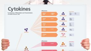 挂图Human Immune Cytokines Infographic of key cytokines for expansion, differentiation and characterization of major immune cell types
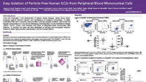 科学海报Easy Isolation of Particle-Free Human ILC2s from Peripheral Blood Mononuclear Cells
 InterviewCaroline Lindemans, MD, PhD How Organoids Provide a Model System for Intestinal Regeneration and Repair
 文献
文献

 沪公网安备31010102008431号