Lerch JK et al. (MAR 2014)
Molecular and Cellular Neuroscience 59 97--105
cJun promotes CNS axon growth
A number of genes regulate regeneration of peripheral axons,but their ability to drive axon growth and regeneration in the central nervous system (CNS) remains largely untested. To address this question we overexpressed eight transcription factors and one small GTPase alone and in pairwise combinations to test whether combinatorial overexpression would have a synergistic impact on CNS neuron neurite growth. The Jun oncogene/signal transducer and activator of transcription 6 (JUN/STAT6) combination increased neurite growth in dissociated cortical neurons and in injured cortical slices. In injured cortical slices,JUN overexpression increased axon growth to a similar extent as JUN and STAT6 together. Interestingly,JUN overexpression was not associated with increased growth associated protein 43 (GAP43) or integrin alpha 7 (ITGA7) expression,though these are predicted transcriptional targets. This study demonstrates that JUN overexpression in cortical neurons stimulates axon growth,but does so independently of changes in expression of genes thought to be critical for JUNs effects on axon growth. We conclude that JUN activity underlies this CNS axonal growth response,and that it is mechanistically distinct from peripheral regeneration responses,in which increases in JUN expression coincide with increases in GAP43 expression.
View Publication
文献
Lee K et al. (JAN 2013)
Neuron 77 1 99--114
Mossy Fiber-CA3 Synapses Mediate Homeostatic Plasticity in Mature Hippocampal Neurons
Network activity homeostatically alters synaptic efficacy to constrain neuronal output. However,it is unclear how such compensatory adaptations coexist with synaptic information storage,especially in established networks. Here,we report that in mature hippocampal neurons in vitro,network activity preferentially regulated excitatory synapses within the proximal dendrites of CA3 neurons. These homeostatic synapses exhibited morphological,functional,and molecular signatures of the specialized contacts between mossy fibers of dentate granule cells and thorny excrescences (TEs) of CA3 pyramidal neurons. In vivo TEs were also selectively and bidirectionally altered by chronic activity changes. TE formation required presynaptic synaptoporin and was suppressed by the activity-inducible kinase,Plk2. These results implicate the mossy fiber-TE synapse as an independently tunable gain control locus that permits efficacious homeostatic adjustment of mossy fiber-CA3 synapses,while preserving synaptic weights that may encode information elsewhere within the mature hippocampal circuit.
View Publication
文献
Leal G et al. (OCT 2014)
PLoS ONE 9 10 e108175
Neuronal Activity Induces Synaptic Delivery of hnRNP A2/B1 by a BDNF-Dependent Mechanism in Cultured Hippocampal Neurons
Dendritic protein synthesis plays a critical role in several forms of synaptic plasticity,including BDNF (brain-derived neurotrophic factor)-mediated long-term synaptic potentiation (LTP). Dendritic transcripts are typically transported in a repressed state as components of large ribonucleoprotein complexes,and then translated upon stimulation at,or in the vicinity,of activated synapses. Heterogeneous nuclear ribonucleoprotein A2/B1 (hnRNP A2/B1) is a trans-acting factor involved in dendritic mRNA trafficking,but how the distribution of the protein in dendrites is regulated has not been characterized. Here we found that a fraction of hnRNP A2/B1 is present at the synapse under resting conditions in cultured hippocampal neurons. Accordingly,this ribonucleoprotein was detected in free mRNP,monosomal,and polyribosomal fractions obtained from synaptoneurosomes. Neuronal activity and BDNF treatment increased hnRNP A2/B1 protein levels in the cell body and dendritic compartments,and induced the delivery of this protein to synaptic sites. The activity-dependent accumulation of hnRNP A2/B1 at the synapse required,at least in part,the activation of TrkB receptors,presumably by BDNF. This neurotrophin also upregulated the hnRNP A2/B1 mRNA in the soma but was without effect on the abundance of neuritic hnRNP A2/B1 transcripts. These results show that the distribution of hnRNP A2/B1 is regulated by BDNF and by neuronal activity,an effect that may have a role in BDNF-induced synaptic plasticity events.
View Publication
On-demand optogenetic activation of human stem-cell-derived neurons
The widespread application of human stem-cell-derived neurons for functional studies is impeded by complicated differentiation protocols,immaturity,and deficient optogene expression as stem cells frequently lose transgene expression over time. Here we report a simple but precise Cre-loxP-based strategy for generating conditional,and thereby stable,optogenetic human stem-cell lines. These cells can be easily and efficiently differentiated into functional neurons,and optogene expression can be triggered by administering Cre protein to the cultures. This conditional expression system may be applied to stem-cell-derived neurons whenever timed transgene expression could help to overcome silencing at the stem-cell level.
View Publication
文献
Kayama T et al. (JAN 2018)
Biochemical and Biophysical Research Communications 495 1 1028--1033
Temporally coordinated spiking activity of human induced pluripotent stem cell-derived neurons co-cultured with astrocytes
In culture conditions,human induced-pluripotent stem cells (hiPSC)-derived neurons form synaptic connections with other cells and establish neuronal networks,which are expected to be an in vitro model system for drug discovery screening and toxicity testing. While early studies demonstrated effects of co-culture of hiPSC-derived neurons with astroglial cells on survival and maturation of hiPSC-derived neurons,the population spiking patterns of such hiPSC-derived neurons have not been fully characterized. In this study,we analyzed temporal spiking patterns of hiPSC-derived neurons recorded by a multi-electrode array system. We discovered that specific sets of hiPSC-derived neurons co-cultured with astrocytes showed more frequent and highly coherent non-random synchronized spike trains and more dynamic changes in overall spike patterns over time. These temporally coordinated spiking patterns are physiological signs of organized circuits of hiPSC-derived neurons and suggest benefits of co-culture of hiPSC-derived neurons with astrocytes.
View Publication
文献
Katikireddy KR et al. (OCT 2016)
The American Journal of Pathology 186 10 2736--2750
Existence of Neural CrestDerived Progenitor Cells in Normal and Fuchs Endothelial Dystrophy Corneal Endothelium
Human corneal endothelial cells are derived from neural crest and because of postmitotic arrest lack competence to repair cell loss from trauma,aging,and degenerative disorders such as Fuchs endothelial corneal dystrophy (FECD). Herein,we identified a rapidly proliferating subpopulation of cells from the corneal endothelium of adult normal and FECD donors that exhibited features of neural crest-derived progenitor (NCDP) cells by showing absence of senescence with passaging,propensity to form spheres,and increased colony forming efficacy compared with the primary cells. The collective expression of stem cell-related genes SOX2,OCT4,LGR5,TP63 (p63),as well as neural crest marker genes PSIP1 (p75(NTR)),PAX3,SOX9,AP2B1 (AP-2β),and NES,generated a phenotypic footprint of endothelial NCDPs. NCDPs displayed multipotency by differentiating into microtubule-associated protein 2,β-III tubulin,and glial fibrillary acidic protein positive neurons and into p75(NTR)-positive human corneal endothelial cells that exhibited transendothelial resistance of functional endothelium. In conclusion,we found that mitotically incompetent ocular tissue cells contain adult NCDPs that exhibit a profile of transcription factors regulating multipotency and neural crest progenitor characteristics. Identification of normal NCDPs in FECD-affected endothelium holds promise for potential autologous cell therapies.
View Publication
文献
Jessick VJ et al. ( 2013)
International journal of physiology,pathophysiology and pharmacology 5 4 216--27
Investigating the role of the actin regulating complex ARP2/3 in rapid ischemic tolerance induced neuro-protection.
Neuronal morphology is highly sensitive to ischemia,although some re-organization may promote neuroprotection. In this study we investigate the role of actin regulating proteins (ARP2,ARP3 and WAVE-1) and their role in rapid ischemic tolerance. Using an established in vitro model of rapid ischemic tolerance,we show that WAVE-1 protein levels are stabilized following brief tolerance inducing ischemia (preconditioning). The stabilization appears to be due to a reduction in the ubiquitination of WAVE-1. Levels of ARP2,ARP3 and N-WASP were not affected by ischemic preconditioning. Immunocytochemical studies show a relocalization of ARP2 and ARP3 proteins in neurons following preconditioning ischemia,as well as a re-organization of actin. Blocking the protein kinase CK2 using emodin blocks ischemic tolerance,and our data suggests CK2 binds to WAVE-1 in neurons. We observe an increase in binding of the ARP2 subunit with WAVE-1. The neuroprotection observed following preconditioning is inhibited when cells are transduced with an N-WASP CA domain that blocks the activation of ARP2/3. Together these data show that ischemia affects actin regulating enzymes,and that the ARP2/3 pathway plays a role in rapid ischemic tolerance induced neuroprotection.
View Publication
文献
Jackson TC et al. (FEB 2018)
Experimental Neurology 300 232--246
BrainPhys increases neurofilament levels in CNS cultures, and facilitates investigation of axonal damage after a mechanical stretch-injury in vitro
Neurobasal®/B27 is a gold standard culture media used to study primary neurons in vitro. An alternative media (BrainPhys®/SM1) was recently developed which robustly enhances neuronal activity vs. Neurobasal® or DMEM. To the best of our knowledge BrainPhys® has not been explored in the setting of neuronal injury. Here we characterized the utility of BrainPhys® in a model of in vitro mechanical-stretch injury. METHODS/RESULTSPrimary rat cortical neurons were maintained in classic Neurobasal®,or sequentially maintained in Neurocult® followed by BrainPhys® (hereafter simply referred to as BrainPhys® maintained neurons?). The levels of axonal markers and proteins involved in neurotransmission were compared on day in vitro 10 (DIV10). BrainPhys® maintained neurons had higher levels of GluN2B,GluR1,Neurofilament light/heavy chain (NF-L & NF-H),and protein phosphatase 2 A (PP2A) vs. neurons in Neurobasal®. Mechanical stretch-injury (50ms/54% biaxial stretch) to BrainPhys® maintained neurons modestly (albeit significantly) increased 24h lactate dehydrogenase (LDH) levels but markedly decreased axonal NF-L levels post-injury vs. uninjured controls or neurons given a milder 38% stretch-injury. Furthermore,two 54% stretch-injuries (in tandem) exacerbated 24h LDH release,increased α-spectrin breakdown products (SBDPs),and decreased Tau levels. Also,BrainPhys® maintained cultures had decreased markers of cell damage 24h after a single 54% stretch-injury vs. neurons in Neurobasal®. Finally,we tested the hypothesis that lentivirus mediated overexpression of the pro-death protein RBM5 exacerbates neuronal and/or axonal injury in primary CNS cultures. RBM5 overexpression vs. empty-vector controls increased 24h LDH release,and SBDP levels,after a single 54% stretch-injury but did not affect NF-L levels or Tau. CONCLUSIONBrainPhys® is a promising new reagent which facilities the investigation of molecular targets involved in axonal and/or neuronal injury in vitro.
View Publication
文献
Aflaki E et al. (JUL 2016)
Journal of Neuroscience 36 28 7441--7452
A New Glucocerebrosidase Chaperone Reduces -Synuclein and Glycolipid Levels in iPSC-Derived Dopaminergic Neurons from Patients with Gaucher Disease and Parkinsonism
UNLABELLED Among the known genetic risk factors for Parkinson disease,mutations in GBA1,the gene responsible for the lysosomal disorder Gaucher disease,are the most common. This genetic link has directed attention to the role of the lysosome in the pathogenesis of parkinsonism. To study how glucocerebrosidase impacts parkinsonism and to evaluate new therapeutics,we generated induced human pluripotent stem cells from four patients with Type 1 (non-neuronopathic) Gaucher disease,two with and two without parkinsonism,and one patient with Type 2 (acute neuronopathic) Gaucher disease,and differentiated them into macrophages and dopaminergic neurons. These cells exhibited decreased glucocerebrosidase activity and stored the glycolipid substrates glucosylceramide and glucosylsphingosine,demonstrating their similarity to patients with Gaucher disease. Dopaminergic neurons from patients with Type 2 and Type 1 Gaucher disease with parkinsonism had reduced dopamine storage and dopamine transporter reuptake. Levels of α-synuclein,a protein present as aggregates in Parkinson disease and related synucleinopathies,were selectively elevated in neurons from the patients with parkinsonism or Type 2 Gaucher disease. The cells were then treated with NCGC607,a small-molecule noninhibitory chaperone of glucocerebrosidase identified by high-throughput screening and medicinal chemistry structure optimization. This compound successfully chaperoned the mutant enzyme,restored glucocerebrosidase activity and protein levels,and reduced glycolipid storage in both iPSC-derived macrophages and dopaminergic neurons,indicating its potential for treating neuronopathic Gaucher disease. In addition,NCGC607 reduced α-synuclein levels in dopaminergic neurons from the patients with parkinsonism,suggesting that noninhibitory small-molecule chaperones of glucocerebrosidase may prove useful for the treatment of Parkinson disease. SIGNIFICANCE STATEMENT Because GBA1 mutations are the most common genetic risk factor for Parkinson disease,dopaminergic neurons were generated from iPSC lines derived from patients with Gaucher disease with and without parkinsonism. These cells exhibit deficient enzymatic activity,reduced lysosomal glucocerebrosidase levels,and storage of glucosylceramide and glucosylsphingosine. Lines generated from the patients with parkinsonism demonstrated elevated levels of α-synuclein. To reverse the observed phenotype,the neurons were treated with a novel noninhibitory glucocerebrosidase chaperone,which successfully restored glucocerebrosidase activity and protein levels and reduced glycolipid storage. In addition,the small-molecule chaperone reduced α-synuclein levels in dopaminergic neurons,indicating that chaperoning glucocerebrosidase to the lysosome may provide a novel therapeutic strategy for both Parkinson disease and neuronopathic forms of Gaucher disease.
View Publication
Xu X et al. (MAR 2017)
Stem Cell Reports 8 3 619--633
Reversal of Phenotypic Abnormalities by CRISPR/Cas9-Mediated Gene Correction in Huntington Disease Patient-Derived Induced Pluripotent Stem Cells
Huntington disease (HD) is a dominant neurodegenerative disorder caused by a CAG repeat expansion in HTT. Here we report correction of HD human induced pluripotent stem cells (hiPSCs) using a CRISPR-Cas9 and piggyBac transposon-based approach. We show that both HD and corrected isogenic hiPSCs can be differentiated into excitable,synaptically active forebrain neurons. We further demonstrate that phenotypic abnormalities in HD hiPSC-derived neural cells,including impaired neural rosette formation,increased susceptibility to growth factor withdrawal,and deficits in mitochondrial respiration,are rescued in isogenic controls. Importantly,using genome-wide expression analysis,we show that a number of apparent gene expression differences detected between HD and non-related healthy control lines are absent between HD and corrected lines,suggesting that these differences are likely related to genetic background rather than HD-specific effects. Our study demonstrates correction of HD hiPSCs and associated phenotypic abnormalities,and the importance of isogenic controls for disease modeling using hiPSCs.
View Publication

 EasySep™小鼠TIL(CD45)正选试剂盒


 文献
文献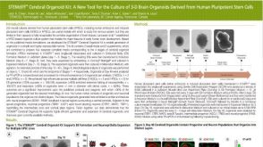 科学海报STEMdiff Cerebral Organoid Kit: A New Tool for the Culture of 3D Brain Organoids Derived from Human Pluripotent Stem Cells
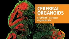

 沪公网安备31010102008431号