A. Sehgal et al. (MAR 2018)
Nature communications 9 1 1272
The role of CSF1R-dependent macrophages in control of the intestinal stem-cell niche.
Colony-stimulating factor 1 (CSF1) controls the growth and differentiation of macrophages.CSF1R signaling has been implicated in the maintenance of the intestinal stem cell niche and differentiation of Paneth cells,but evidence of expression of CSF1R within the crypt is equivocal. Here we show that CSF1R-dependent macrophages influence intestinal epithelial differentiation and homeostasis. In the intestinal lamina propria CSF1R mRNA expression is restricted to macrophages which are intimately associated with the crypt epithelium,and is undetectable in Paneth cells. Macrophage ablation following CSF1R blockade affects Paneth cell differentiation and leads to a reduction of Lgr5+ intestinal stem cells. The disturbances to the crypt caused by macrophage depletion adversely affect the subsequent differentiation of intestinal epithelial cell lineages. Goblet cell density is enhanced,whereas the development of M cells in Peyer's patches is impeded. We suggest that modification of the phenotype or abundance of macrophages in the gut wall alters the development of the intestinal epithelium and the ability to sample gut antigens.
View Publication
文献
D. R. McHugh et al. ( 2018)
PloS one 13 6 e0199573
A G542X cystic fibrosis mouse model for examining nonsense mutation directed therapies.
Nonsense mutations are present in 10{\%} of patients with CF,produce a premature termination codon in CFTR mRNA causing early termination of translation,and lead to lack of CFTR function. There are no currently available animal models which contain a nonsense mutation in the endogenous Cftr locus that can be utilized to test nonsense mutation therapies. In this study,we create a CF mouse model carrying the G542X nonsense mutation in Cftr using CRISPR/Cas9 gene editing. The G542X mouse model has reduced Cftr mRNA levels,demonstrates absence of CFTR function,and displays characteristic manifestations of CF mice such as reduced growth and intestinal obstruction. Importantly,CFTR restoration is observed in G542X intestinal organoids treated with G418,an aminoglycoside with translational readthrough capabilities. The G542X mouse model provides an invaluable resource for the identification of potential therapies of CF nonsense mutations as well as the assessment of in vivo effectiveness of these potential therapies targeting nonsense mutations.
View Publication
文献
C. L. Kraft et al. (NOV 2017)
Oncotarget 8 61 102923--102933
GUCY2C maintains intestinal LGR5+stem cells by opposing ER stress.
Long-lived multipotent stem cells (ISCs) at the base of intestinal crypts adjust their phenotypes to accommodate normal maintenance and post-injury regeneration of the epithelium. Their long life,lineage plasticity,and proliferative potential underlie the necessity for tight homeostatic regulation of the ISC compartment. In that context,the guanylate cyclase C (GUCY2C) receptor and its paracrine ligands regulate intestinal epithelial homeostasis,including proliferation,lineage commitment,and DNA damage repair. However,a role for this axis in maintaining ISCs remains unknown. Transgenic mice enabling analysis of ISCs (Lgr5-GFP) in the context of GUCY2C elimination (Gucy2c -/- ) were combined with immunodetection techniques and pharmacological treatments to define the role of the GUCY2C signaling axis in supporting ISCs. ISCs were reduced inGucy2c -/- mice,associated with loss of active Lgr5+cells but a reciprocal increase in reserve Bmi1+cells. GUCY2C was expressed in crypt base Lgr5+cells in which it mediates canonical cyclic (c) GMP-dependent signaling. Endoplasmic reticulum (ER) stress,typically absent from ISCs,was elevated throughout the crypt base inGucy2c -/- mice. The chemical chaperone tauroursodeoxycholic acid resolved this ER stress and restored the balance of ISCs,an effect mimicked by the GUCY2C effector 8Br-cGMP. Reduced ISCs inGucy2c -/- mice was associated with greater epithelial injury and impaired regeneration following sub-lethal doses of irradiation. These observations suggest that GUCY2C provides homeostatic signals that modulate ER stress and cell vulnerability as part of the machinery contributing to the integrity of ISCs.
View Publication
文献
S. Ihara et al. (JUN 2018)
Journal of Crohn's & colitis
Adhesive interactions between Mononuclear Phagocytes and Intestinal Epithelium Perturb Normal Epithelial Differentiation and Serve as a Therapeutic Target in Inflammatory Bowel Disease.
Background and Aims Disturbance of intestinal homeostasis is associated with the development of inflammatory bowel disease (IBD),and TGF-beta$ signaling impairment in mononuclear phagocytes (MPs) causes murine colitis with goblet cell depletion. Here,we examined an organoid-MP co-culture system to study the role of MPs in intestinal epithelial differentiation and homeostasis. Methods Intestinal organoids were co-cultured with lamina propria leukocytes and bone marrow-derived dendritic cells (BMDCs) from CD11c-cre Tgfbr2fl/fl mice. Organoid-MP adhesive interactions were evaluated by microscopy,RT-PCR,and flow cytometry. Murine colitis models (dextran sodium sulphate (DSS),CD11c-cre Tgfbr2fl/fl,T-cell-transfer) were used for histological and immunohistochemical analysis. Anti-E-cadherin antibody treatment or CD11c+-cell-specific CDH1 gene deletion were performed for E-cadherin neutralization or knockout. Colonic biopsies from patients with ulcerative colitis were analyzed by flow cytometry. Results Intestinal organoids co-cultured with CD11c+ lamina propria leukocytes or BMDCs from CD11c-cre Tgfbr2fl/fl mice showed morphological changes and goblet cell depletion with Notch signal activation,analogous to CD11c-cre Tgfbr2fl/fl colitis. E-cadherin was upregulated in CD11c+ MPs,especially CX3CR1+CCR2+ monocytes,of CD11c-cre Tgfbr2fl/fl mice. E-cadherin-mediated BMDC adhesion promoted Notch activation and cystic changes in organoids. Anti-E-cadherin antibody treatment attenuated colitis in CD11c-cre Tgfbr2fl/fl and T-cell-transferred mice. In addition,E-cadherin deletion in CD11c+ cells attenuated colitis in both CD11c-cre Tgfbr2fl/fl and DSS-treated mice. In patients with ulcerative colitis,E-cadherin expressed by intestinal CD11c+ leukocytes was enhanced compared with that in healthy controls. Conclusions E-cadherin-mediated MP-epithelium adhesion is associated with the development of colitis,and blocking these adhesions may have therapeutic potential for IBD.
View Publication
文献
K. Huang et al. (MAY 2018)
Pediatric research 83 5 1031--1040
Targeting the PXR-TLR4 signaling pathway to reduce intestinal inflammation in an experimental model of necrotizing enterocolitis.
BackgroundThere is substantial evidence that signaling through Toll-like receptor 4 (TLR4) contributes to the pathogenesis of necrotizing enterocolitis (NEC). Pregnane X receptor (PXR),a xenobiotic sensor and signaling intermediate for certain host-bacterial metabolites,has been shown to negatively regulate TLR4 signaling. Here we investigated the relationship between PXR and TLR4 in the developing murine intestine and explored the capacity of PXR to modulate inflammatory pathways involved in experimental NEC.MethodsWild-type and PXR-/- mice were studied at various time points of development in an experimental model of NEC. In addition,we studied the ability of the secondary bile acid lithocholic acid (LCA),a known PXR agonist in liver,to activate intestinal PXR and reduce NEC-related intestinal inflammation.ResultsWe found a reciprocal relationship between the developmental expression of PXR and TLR4 in wild-type murine intestine,with PXR acting to reduce TLR4 expression by decreasing TLR4 mRNA stability. In addition,PXR-/- mice exhibited a remarkably heightened severity of disease in experimental NEC. Moreover,LCA attenuated intestinal proinflammatory responses in the early stages of experimental NEC.ConclusionThese findings provide proactive insights into the regulation of TLR4 in the developing intestine. Targeting PXR may be a novel approach for NEC prevention.
View Publication
文献
M. D. Hu et al. (JUL 2018)
Journal of immunology (Baltimore,Md. : 1950) 201 2 747--756
Epithelial IL-15 Is a Critical Regulator of gamma$delta$ Intraepithelial Lymphocyte Motility within the Intestinal Mucosa.
Intraepithelial lymphocytes (IELs) expressing the gamma$delta$ TCR (gamma$delta$ IELs) provide continuous surveillance of the intestinal epithelium. However,the mechanisms regulating the basal motility of these cells within the epithelial compartment have not been well defined. We investigated whether IL-15 contributes to gamma$delta$ IEL localization and migratory behavior in addition to its role in IEL differentiation and survival. Using advanced live cell imaging techniques in mice,we find that compartmentalized overexpression of IL-15 in the lamina propria shifts the distribution of gamma$delta$ T cells from the epithelial compartment to the lamina propria. This mislocalization could be rescued by epithelial IL-15 overexpression,indicating that epithelial IL-15 is essential for gamma$delta$ IEL migration into the epithelium. Furthermore,in vitro analyses demonstrated that exogenous IL-15 stimulates gamma$delta$ IEL migration into cultured epithelial monolayers,and inhibition of IL-2Rbeta$ significantly attenuates the basal motility of these cells. Intravital microscopy showed that impaired IL-2Rbeta$ signaling induced gamma$delta$ IEL idling within the lateral intercellular space,which resulted in increased early pathogen invasion. Similarly,the redistribution of gamma$delta$ T cells to the lamina propria due to local IL-15 overproduction also enhanced bacterial translocation. These findings thus reveal a novel role for IL-15 in mediating gamma$delta$ T cell localization within the intestinal mucosa and regulating gamma$delta$ IEL motility and patrolling behavior as a critical component of host defense.
View Publication
文献
R. M. Eichenberger et al. ( 2018)
Journal of extracellular vesicles 7 1 1428004
Characterization ofTrichuris murissecreted proteins and extracellular vesicles provides new insights into host-parasite communication.
Whipworms are parasitic nematodes that live in the gut of more than 500 million people worldwide. Owing to the difficulty in obtaining parasite material,the mouse whipwormTrichuris murishas been extensively used as a model to study human whipworm infections. These nematodes secrete a multitude of compounds that interact with host tissues where they orchestrate a parasitic existence. Herein we provide the first comprehensive characterization of the excretory/secretory products ofT. muris. We identify 148 proteins secreted byT. murisand show for the first time that the mouse whipworm secretes exosome-like extracellular vesicles (EVs) that can interact with host cells. We use an Optiprep{\textregistered} gradient to purify the EVs,highlighting the suitability of this method for purifying EVs secreted by a parasitic nematode. We also characterize the proteomic and genomic content of the EVs,identifying {\textgreater}350 proteins,56 miRNAs (22 novel) and 475 full-length mRNA transcripts mapping toT. murisgene models. Many of the miRNAs putatively mapped to mouse genes are involved in regulation of inflammation,implying a role in parasite-driven immunomodulation. In addition,for the first time to our knowledge,colonic organoids have been used to demonstrate the internalization of parasite EVs by host cells. Understanding how parasites interact with their host is crucial to develop new control measures. This first characterization of the proteins and EVs secreted byT. murisprovides important information on whipworm-host communication and forms the basis for future studies.
View Publication
文献
E. A. Davis et al. (JUN 2018)
Physiological reports 6 12 e13745
Evidence for a direct effect of the autonomic nervous system on intestinal epithelial stem cell proliferation.
The sympathetic (SNS) and parasympathetic (PNS) branches of the autonomic nervous system have been implicated in the modulation of the renewal of many tissues,including the intestinal epithelium. However,it is not known whether these mechanisms are direct,requiring an interaction between autonomic neurotransmitters and receptors on proliferating epithelial cells. To evaluate the existence of a molecular framework for a direct effect of the SNS or PNS on intestinal epithelial renewal,we measured gene expression for the main autonomic neurotransmitter receptors in this tissue. We separately evaluated intestinal epithelial regions comprised of the stem,progenitor,and mature cells,which allowed us to investigate the distinct contributions of each cell population to this proposed autonomic effect. Notably,we found that the stem cells expressed the receptors for the SNS-associated alpha2A adrenoreceptor and the PNS-associated muscarinic acetylcholine receptors (M1 and M3). In a separate experiment,we found that the application of norepinephrine or acetylcholine decreases the expression of cyclin D1,a gene necessary for cell cycle progression,in intestinal epithelial organoids compared with controls (P {\textless} 0.05). Together,these results provide evidence of a direct mechanism for the autonomic nervous system influence on intestinal epithelial stem cell proliferation.
View Publication
文献
Y. Bhattarai et al. (JUN 2018)
Cell host & microbe 23 6 775--785.e5
Gut Microbiota-Produced Tryptamine Activates an Epithelial G-Protein-Coupled Receptor to Increase Colonic Secretion.
Tryptamine,a tryptophan-derived monoamine similar to 5-hydroxytryptamine (5-HT),is produced by gut bacteria and is abundant in human and rodent feces. However,the physiologic effect of tryptamine in the gastrointestinal (GI) tract remains unknown. Here,we show that the biological effects of tryptamine are mediated through the 5-HT4 receptor (5-HT4R),a G-protein-coupled receptor (GPCR) uniquely expressed in the colonic epithelium. Tryptamine increases both ionic flux across the colonic epithelium and fluid secretion in colonoids from germ-free (GF) and humanized (ex-GF colonized with human stool) mice,consistent with increased intestinal secretion. The secretory effect of tryptamine is dependent on 5-HT4R activation and is blocked by 5-HT4R antagonist and absent in 5-HT4R-/- mice. GF mice colonized by Bacteroides thetaiotaomicron engineered to produce tryptamine exhibit accelerated GI transit. Our study demonstrates an aspect of host physiology under control of a bacterial metabolite that can be exploited as a therapeutic modality. VIDEO ABSTRACT.
View Publication

 EasySep™小鼠TIL(CD45)正选试剂盒


 文献
文献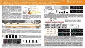 科学海报Highly Efficient Differentiation of Human Pluripotent Stem Cells into Long-term Expandable “Mini-gut” Organoids

 沪公网安备31010102008431号