Targeting placental growth factor/neuropilin 1 pathway inhibits growth and spread of medulloblastoma.
Medulloblastoma is the most common pediatric malignant brain tumor. Although current therapies improve survival,these regimens are highly toxic and are associated with significant morbidity. Here,we report that placental growth factor (PlGF) is expressed in the majority of medulloblastomas,independent of their subtype. Moreover,high expression of PlGF receptor neuropilin 1 (Nrp1) correlates with poor overall survival in patients. We demonstrate that PlGF and Nrp1 are required for the growth and spread of medulloblastoma: PlGF/Nrp1 blockade results in direct antitumor effects in vivo,resulting in medulloblastoma regression,decreased metastasis,and increased mouse survival. We reveal that PlGF is produced in the cerebellar stroma via tumor-derived Sonic hedgehog (Shh) and show that PlGF acts through Nrp1-and not vascular endothelial growth factor receptor 1-to promote tumor cell survival. This critical tumor-stroma interaction-mediated by Shh,PlGF,and Nrp1 across medulloblastoma subtypes-supports the development of therapies targeting PlGF/Nrp1 pathway.
View Publication
产品号#:
05700
05701
05702
产品名:
NeuroCult™ 基础培养基(小鼠和大鼠)
NeuroCult™ 扩增添加物(小鼠和大鼠)
NeuroCult™扩增试剂盒(小鼠和大鼠)
Xu G et al. (MAY 2013)
Neuroscience 238 195--208
Functional analysis of platelet-derived growth factor receptor-β in neural stem/progenitor cells
Activation of neural stem/progenitor cells (NSPCs) is a potential therapeutic strategy of neurological disorders. In this study,NSPCs of subventricular zone were isolated and cultured from platelet-derived growth factor-β-receptor-knockout (PDGFR-β(-/-)) mice of postnatal day 1 (P1) and P28,and the roles of PDGFR-β were examined in these cells. In PDGFR-β-preserving control NSPCs,stem cell activities,such as numbers and diameters of secondary neurospheres,cell proliferation and survival rates,were significantly higher in P1 NSPCs than those in P28 NSPCs. In PDGFR-β(-/-) NSPCs,most of these parameters were decreased as compared with age-matched controls. Among them,the decrease of secondary neurosphere formation was most striking in P1 and P28 PDGFR-β(-/-) NSPCs and in P28 control NSPCs as compared with P1 control NSPCs. PCR-array and following quantitative real-time PCR (qRT-PCR) analyses demonstrated that expressions of fibroblast growth factor-2 (FGF2) and exons IV-IX of brain-derived neurotrophic factor (BDNF) were decreased,and noggin was increased in P1 PDGFR-β(-/-) as compared with P1 controls. Addition of BDNF rescued the number and diameter of secondary neurospheres in P1 PDGFR-β(-/-) NSPCs to similar levels as controls. The expressions of PDGFs and PDGFRs in control NSPCs were increased along with the differentiation-induction,where phosphorylated PDGFR-β was co-localized with neuronal and astrocyte differentiation markers. In controls,the neuronal differentiation was decreased,and the glial differentiation was increased from P1 to P28 NSPCs. Compared with P1 controls,neuronal differentiation was reduced in P1 PDGFR-β(-/-) NSPCs,whereas glial differentiation was comparable between the two genotypes. These results suggest that PDGFR-β signaling is important for the self-renewal and multipotency of NSPCs,particularly in neonatal NSPCs. BDNF,FGF2,and noggin may be involved in the effects of PDGFR-β signaling in these cells. Accordingly,the activation of PDGFR-β in NSPCs may be a novel therapeutic strategy of neurological diseases.
View Publication
Ishii Y et al. (MAR 2008)
Molecular and cellular neurosciences 37 3 507--18
Characterization of neuroprogenitor cells expressing the PDGF beta-receptor within the subventricular zone of postnatal mice.
We report a considerable number of cells in the ventricular and the subventricular zones (SVZ) of newborn mice to stain positive for the PDGF beta-receptor (PDGFRB). Many of them also stained for nestin and/or GFAP but less frequently for the neuroblast marker doublecortin and for the mitotic marker Ki-67. The SVZ of mice with nestin-Cre conditional deletion of PDGFRB expressed the receptor only on blood vessels and was devoid of any morphological abnormality. PDGFRB(-/-) neurospheres showed a higher rate of apoptosis without any significant decrease in proliferation. They demonstrated reduced capacities of migration and neuronal differentiation in response to not only PDGF-BB but also bFGF. Furthermore,the PDGFR kinase inhibitor STI571 blocked the effects of bFGF in control neurosphere cultures. bFGF increased the activity of the PDGFRB promoter as well as the expression and phosphorylation of PDGFRB. These results suggest the presence of the signaling convergence between PDGF and FGF. PDGFRB is needed for survival,and the effects of bFGF in migration and neural differentiation of the cells may be potentiated by induction of PDGFRB.
View Publication
产品号#:
05700
05701
05702
产品名:
NeuroCult™ 基础培养基(小鼠和大鼠)
NeuroCult™ 扩增添加物(小鼠和大鼠)
NeuroCult™扩增试剂盒(小鼠和大鼠)
Chua SJ et al. (FEB 2009)
Biochemical and biophysical research communications 379 2 217--21
Neural progenitors, neurons and oligodendrocytes from human umbilical cord blood cells in a serum-free, feeder-free cell culture.
We have previously demonstrated that lineage negative cells (Lin(neg)) from umbilical cord blood (UCB) develop into multipotent cells capable of differentiation into bone,muscle,endothelial and neural cells. The objective of this study was to determine the optimal conditions required for Lin(neg) UCB cells to differentiate into neuronal cells and oligodendrocytes. We demonstrate that early neural stage markers (nestin,neurofilament,A2B5 and Sox2) are expressed in Lin(neg) cells cultured in FGF4,SCF,Flt3-ligand reprogramming culture media followed by the early macroglial cell marker O4. Early stage oligodendrocyte markers CNPase,GalC,Olig2 and the late-stage marker MOSP are observed,as is the Schwann cell marker PMP22. In summary,Lin(neg) UCB cells,when appropriately cultured,are able to exhibit characteristics of neuronal and macroglial cells that can specifically differentiate into oligodendrocytes and Schwann cells and express proteins associated with myelin production after in vitro differentiation.
View Publication
产品号#:
09600
09650
产品名:
StemSpan™ SFEM
StemSpan™ SFEM
Wakimoto H et al. (APR 2009)
Cancer research 69 8 3472--81
Human glioblastoma-derived cancer stem cells: establishment of invasive glioma models and treatment with oncolytic herpes simplex virus vectors.
Glioblastoma,the most malignant type of primary brain tumor,is one of the solid cancers where cancer stem cells have been isolated,and studies have suggested resistance of those cells to chemotherapy and radiotherapy. Here,we report the establishment of CSC-enriched cultures derived from human glioblastoma specimens. They grew as neurospheres in serum-free medium with epidermal growth factor and fibroblast growth factor 2,varied in the level of CD133 expression and very efficiently formed highly invasive and/or vascular tumors upon intracerebral implantation into immunodeficient mice. As a novel therapeutic strategy for glioblastoma-derived cancer stem-like cells (GBM-SC),we have tested oncolytic herpes simplex virus (oHSV) vectors. We show that although ICP6 (UL39)-deleted mutants kill GBM-SCs as efficiently as wild-type HSV,the deletion of gamma34.5 significantly attenuated the vectors due to poor replication. However,this was significantly reversed by the additional deletion of alpha47. Infection with oHSV G47Delta (ICP6(-),gamma34.5(-),alpha47(-)) not only killed GBM-SCs but also inhibited their self-renewal as evidenced by the inability of viable cells to form secondary tumor spheres. Importantly,despite the highly invasive nature of the intracerebral tumors generated by GBM-SCs,intratumoral injection of G47Delta significantly prolonged survival. These results for the first time show the efficacy of oHSV against human GBM-SCs,and correlate this cytotoxic property with specific oHSV mutations. This is important for designing new oHSV vectors and clinical trials. Moreover,the new glioma models described in this study provide powerful tools for testing experimental therapeutics and studying invasion and angiogenesis.
View Publication
产品号#:
05707
产品名:
NeuroCult™化学解离试剂盒(小鼠)
Kronenberg G et al. (MAR 2010)
The Journal of neuroscience : the official journal of the Society for Neuroscience 30 9 3419--31
Impact of actin filament stabilization on adult hippocampal and olfactory bulb neurogenesis.
Rearrangement of the actin cytoskeleton is essential for dynamic cellular processes. Decreased actin turnover and rigidity of cytoskeletal structures have been associated with aging and cell death. Gelsolin is a Ca(2+)-activated actin-severing protein that is widely expressed throughout the adult mammalian brain. Here,we used gelsolin-deficient (Gsn(-/-)) mice as a model system for actin filament stabilization. In Gsn(-/-) mice,emigration of newly generated cells from the subventricular zone into the olfactory bulb was slowed. In vitro,gelsolin deficiency did not affect proliferation or neuronal differentiation of adult neural progenitors cells (NPCs) but resulted in retarded migration. Surprisingly,hippocampal neurogenesis was robustly induced by gelsolin deficiency. The ability of NPCs to intrinsically sense excitatory activity and thereby implement coupling between network activity and neurogenesis has recently been established. Depolarization-induced [Ca(2+)](i) increases and exocytotic neurotransmitter release were enhanced in Gsn(-/-) synaptosomes. Importantly,treatment of Gsn(-/-) synaptosomes with mycotoxin cytochalasin D,which,like gelsolin,produces actin disassembly,decreased enhanced Ca(2+) influx and subsequent exocytotic norepinephrine release to wild-type levels. Similarly,depolarization-induced glutamate release from Gsn(-/-) brain slices was increased. Furthermore,increased hippocampal neurogenesis in Gsn(-/-) mice was associated with a special microenvironment characterized by enhanced density of perfused vessels,increased regional cerebral blood flow,and increased endothelial nitric oxide synthase (NOS-III) expression in hippocampus. Together,reduced filamentous actin turnover in presynaptic terminals causes increased Ca(2+) influx and,subsequently,elevated exocytotic neurotransmitter release acting on neural progenitors. Increased neurogenesis in Gsn(-/-) hippocampus is associated with a special vascular niche for neurogenesis.
View Publication

 EasySep™小鼠TIL(CD45)正选试剂盒


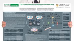 挂图Cell-Reprogramming Technology and Neuroscience Details on human iPSC-derived models of neuropsychiatric and neurodegenerative disorders


 沪公网安备31010102008431号