Erythropoietin (EPO) regulates the proliferation and differentiation of erythroid cells by binding to its specific transmembrane receptor (EPOR). The presence of EPO and its receptor in the CNS suggests a different function for EPO other than erythropoiesis. The purpose of the present study was to examine EPOR expression and the role of EPO in the proliferation of neonatal spinal cord-derived neural progenitor cells. The effect of EPO on cell cycle progression was also examined,as well as the signaling cascades involved in this process. Our results showed that EPOR was present in the neural progenitor cells and EPO significantly enhanced their proliferation. Cell cycle analysis of EPO-treated neural progenitor cells indicated a reduced percentage of cells in G0/G1 phase,whereas the cell proliferation index (S phase plus G2/M phase) was increased. EPO also increased the proportion of 5-bromo-2-deoxyuridine (BrdU)-positive cells. With respect to the cell cycle signaling,we examined the cyclin-dependent kinases D1,D2 and E,and cyclin-dependent kinase inhibitors,p21cip1,p27kip1 and p57kip2. No significant differences were observed in the expression of these transcripts after EPO administration. Interestingly,the anti-apoptotic factors,mcl-1 and bcl-2 were significantly increased twofold. Moreover,these specific effects of EPO were eliminated by incubation of the progenitor cells with anti-EPO neutralizing antibody. Those observations suggested that EPO may play a role in normal spinal cord development by regulating cell proliferation and apoptosis.
View Publication
产品号#:
05771
产品名:
Fernando P et al. (OCT 2005)
FASEB journal : official publication of the Federation of American Societies for Experimental Biology 19 12 1671--3
Neural stem cell differentiation is dependent upon endogenous caspase 3 activity.
Caspase proteases have become the focal point for the development and application of anti-apoptotic therapies in a variety of central nervous system diseases. However,this approach is based on the premise that caspase function is limited to invoking cell death signals. Here,we show that caspase-3 activity is elevated in nonapoptotic differentiating neuronal cell populations. Moreover,peptide inhibition of protease activity effectively inhibits the differentiation process in a cultured neurosphere model. These results implicate caspase-3 activation as a conserved feature of neuronal differentiation and suggest that targeted inhibition of this protease in neural cell populations may have unintended consequences.
View Publication
Sox2 expression defines a heterogeneous population of neurosphere-forming cells in the adult murine brain.
The identification of neural stem cells (NSCs) in situ has been prevented by the inability to identify a marker consistently expressed in all adult NSCs and is thus generally accomplished using the in vitro neurosphere-forming assay. The high-mobility group transcription factor Sox2 is expressed in embryonic neural epithelial stem cells; because these cells are thought to give rise to the adult NSC population,we hypothesized that Sox2 may continue to be expressed in adult NSCs. Using Sox2:EGFP transgenic mice,we show that Sox2 is expressed in neurogenic regions along the rostral-caudal axis of the central nervous system throughout life. Furthermore,all neurospheres derived from these neurogenic regions express Sox2,suggesting that Sox2 is indeed expressed in adult NSCs. We demonstrate that NSCs are heterogeneous within the adult brain,with differing capacities for cell production. In vitro,all neurospheres express Sox2,but the expression of markers common to early progenitor cells within individual neurospheres varies; this heterogeneity of NSCs is mirrored in vivo. For example,both glial fibrillary acidic protein and NG2 are expressed within individual neurospheres,but their expression is mutually exclusive; likewise,these two markers show distinct staining patterns within the Sox2+ regions of the brain's neurogenic regions. Thus,we propose that the expression of Sox2 is a unifying characteristic of NSCs in the adult brain,but that not all NSCs maintain the ability to form all neural cell types in vivo.
View Publication
Guillou L et al. (NOV 2016)
Biophysical journal 111 9 2039--2050
Measuring Cell Viscoelastic Properties Using a Microfluidic Extensional Flow Device.
The quantification of cellular mechanical properties is of tremendous interest in biology and medicine. Recent microfluidic technologies that infer cellular mechanical properties based on analysis of cellular deformations during microchannel traversal have dramatically improved throughput over traditional single-cell rheological tools,yet the extraction of material parameters from these measurements remains quite complex due to challenges such as confinement by channel walls and the domination of complex inertial forces. Here,we describe a simple microfluidic platform that uses hydrodynamic forces at low Reynolds number and low confinement to elongate single cells near the stagnation point of a planar extensional flow. In tandem,we present,to our knowledge,a novel analytical framework that enables determination of cellular viscoelastic properties (stiffness and fluidity) from these measurements. We validated our system and analysis by measuring the stiffness of cross-linked dextran microparticles,which yielded reasonable agreement with previously reported values and our micropipette aspiration measurements. We then measured viscoelastic properties of 3T3 fibroblasts and glioblastoma tumor initiating cells. Our system captures the expected changes in elastic modulus induced in 3T3 fibroblasts and tumor initiating cells in response to agents that soften (cytochalasin D) or stiffen (paraformaldehyde) the cytoskeleton. The simplicity of the device coupled with our analytical model allows straightforward measurement of the viscoelastic properties of cells and soft,spherical objects.
View Publication
产品号#:
05750
05751
产品名:
NeuroCult™ NS-A 基础培养基(人)
NeuroCult™ NS-A 扩增试剂盒(人)
Hackett C et al. ( 2014)
American journal of translational research 6 2 119--28
Transplantation of Fas-deficient or wild-type neural stem/progenitor cells (NPCs) is equally efficient in treating experimental autoimmune encephalomyelitis (EAE).
Studies have shown that neural stem/progenitor cell (NPC) transplantation is beneficial in experimental autoimmune encephalomyelitis (EAE),an established animal model of multiple sclerosis (MS). It is unclear whether NPCs have the ability to integrate into the host CNS to replace lost cells or if their main mechanism of action is via bystander immunomodulation. Understanding the mechanisms by which NPCs exert their beneficial effects as well as exploring methods to increase post-transplantation survival and differentiation is critical to advancing this treatment strategy. Using the EAE model and Fas-deficient (lpr) NPCs,we investigated the effects of altering the Fas system in NPC transplantation therapy. We show that transplantation of NPCs into EAE mice ameliorates clinical symptoms with greater efficacy than sham treatments regardless of cell type (wt or lpr). NPC transplantation via retro-orbital injections significantly decreased inflammatory infiltrates at the acute time point,with a similar trend at the chronic time point. Both wt and lpr NPCs injected into mice with EAE were able to home to sites of CNS inflammation in the periventricular brain and lumbar spinal cord. Both wt and lpr NPCs have the same capacity for inducing apoptosis of Th1 and Th17 cells,and minimal numbers of NPCs entered the CNS. These cells did not express terminal differentiation markers,suggesting that NPCs exert their effects mainly via bystander peripheral immunomodulation.
View Publication
产品号#:
05715
产品名:
NeuroCult™成年中枢神经系统(CNS)组织酶解试剂盒(小鼠和大鼠)
Belkind-Gerson J et al. (JAN 2013)
Neurogastroenterology and motility : the official journal of the European Gastrointestinal Motility Society 25 1 61--9.e7
Nestin-expressing cells in the gut give rise to enteric neurons and glial cells.
BACKGROUND Neuronal stem cells (NSCs) are promising for neurointestinal disease therapy. Although NSCs have been isolated from intestinal musclularis,their presence in mucosa has not been well described. Mucosa-derived NSCs are accessible endoscopically and could be used autologously. Brain-derived Nestin-positive NSCs are important in endogenous repair and plasticity. The aim was to isolate and characterize mucosa-derived NSCs,determine their relationship to Nestin-expressing cells and to demonstrate their capacity to produce neuroglial networks in vitro and in vivo. METHODS Neurospheres were generated from periventricular brain,colonic muscularis (Musc),and mucosa-submucosa (MSM) of mice expressing green fluorescent protein (GFP) controlled by the Nestin promoter (Nestin-GFP). Neuronal stem cells were also grown as adherent colonies from intestinal mucosal organoids. Their differentiation potential was assessed using immunohistochemistry using glial and neuronal markers. Brain and gut-derived neurospheres were transplanted into explants of chick embryonic aneural hindgut to determine their fate. KEY RESULTS Musc- and MSM-derived neurospheres expressed Nestin and gave rise to cells of neuronal,glial,and mesenchymal lineage. Although Nestin expression in tissue was mostly limited to glia co-labelled with glial fibrillary acid protein (GFAP),neurosphere-derived neurons and glia both expressed Nestin in vitro,suggesting that Nestin+/GFAP+ glial cells may give rise to new neurons. Moreover,following transplantation into aneural colon,brain- and gut-derived NSCs were able to differentiate into neurons. CONCLUSIONS & INFERENCES Nestin-expressing intestinal NSCs cells give rise to neurospheres,differentiate into neuronal,glial,and mesenchymal lineages in vitro,generate neurons in vivo and can be isolated from mucosa. Further studies are needed for exploring their potential for treating neuropathies.
View Publication

 EasySep™小鼠TIL(CD45)正选试剂盒


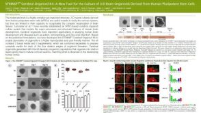 科学海报STEMdiff™ Cerebral Organoid Kit: A New Tool for the Culture of 3D Brain Organoids Derived from hPSCs
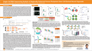 科学海报Single-Cell RNA Sequencing Analysis of Regionally Patterned Human Pluripotent Stem Cell-Derived Neural Organoids


 沪公网安备31010102008431号