Belkind-Gerson J et al. (JAN 2013)
Neurogastroenterology and motility : the official journal of the European Gastrointestinal Motility Society 25 1 61--9.e7
Nestin-expressing cells in the gut give rise to enteric neurons and glial cells.
BACKGROUND Neuronal stem cells (NSCs) are promising for neurointestinal disease therapy. Although NSCs have been isolated from intestinal musclularis,their presence in mucosa has not been well described. Mucosa-derived NSCs are accessible endoscopically and could be used autologously. Brain-derived Nestin-positive NSCs are important in endogenous repair and plasticity. The aim was to isolate and characterize mucosa-derived NSCs,determine their relationship to Nestin-expressing cells and to demonstrate their capacity to produce neuroglial networks in vitro and in vivo. METHODS Neurospheres were generated from periventricular brain,colonic muscularis (Musc),and mucosa-submucosa (MSM) of mice expressing green fluorescent protein (GFP) controlled by the Nestin promoter (Nestin-GFP). Neuronal stem cells were also grown as adherent colonies from intestinal mucosal organoids. Their differentiation potential was assessed using immunohistochemistry using glial and neuronal markers. Brain and gut-derived neurospheres were transplanted into explants of chick embryonic aneural hindgut to determine their fate. KEY RESULTS Musc- and MSM-derived neurospheres expressed Nestin and gave rise to cells of neuronal,glial,and mesenchymal lineage. Although Nestin expression in tissue was mostly limited to glia co-labelled with glial fibrillary acid protein (GFAP),neurosphere-derived neurons and glia both expressed Nestin in vitro,suggesting that Nestin+/GFAP+ glial cells may give rise to new neurons. Moreover,following transplantation into aneural colon,brain- and gut-derived NSCs were able to differentiate into neurons. CONCLUSIONS & INFERENCES Nestin-expressing intestinal NSCs cells give rise to neurospheres,differentiate into neuronal,glial,and mesenchymal lineages in vitro,generate neurons in vivo and can be isolated from mucosa. Further studies are needed for exploring their potential for treating neuropathies.
View Publication
Sox2 expression defines a heterogeneous population of neurosphere-forming cells in the adult murine brain.
The identification of neural stem cells (NSCs) in situ has been prevented by the inability to identify a marker consistently expressed in all adult NSCs and is thus generally accomplished using the in vitro neurosphere-forming assay. The high-mobility group transcription factor Sox2 is expressed in embryonic neural epithelial stem cells; because these cells are thought to give rise to the adult NSC population,we hypothesized that Sox2 may continue to be expressed in adult NSCs. Using Sox2:EGFP transgenic mice,we show that Sox2 is expressed in neurogenic regions along the rostral-caudal axis of the central nervous system throughout life. Furthermore,all neurospheres derived from these neurogenic regions express Sox2,suggesting that Sox2 is indeed expressed in adult NSCs. We demonstrate that NSCs are heterogeneous within the adult brain,with differing capacities for cell production. In vitro,all neurospheres express Sox2,but the expression of markers common to early progenitor cells within individual neurospheres varies; this heterogeneity of NSCs is mirrored in vivo. For example,both glial fibrillary acidic protein and NG2 are expressed within individual neurospheres,but their expression is mutually exclusive; likewise,these two markers show distinct staining patterns within the Sox2+ regions of the brain's neurogenic regions. Thus,we propose that the expression of Sox2 is a unifying characteristic of NSCs in the adult brain,but that not all NSCs maintain the ability to form all neural cell types in vivo.
View Publication
产品号#:
05700
05701
05702
产品名:
NeuroCult™ 基础培养基(小鼠和大鼠)
NeuroCult™ 扩增添加物(小鼠和大鼠)
NeuroCult™扩增试剂盒(小鼠和大鼠)
Fernando P et al. (OCT 2005)
FASEB journal : official publication of the Federation of American Societies for Experimental Biology 19 12 1671--3
Neural stem cell differentiation is dependent upon endogenous caspase 3 activity.
Caspase proteases have become the focal point for the development and application of anti-apoptotic therapies in a variety of central nervous system diseases. However,this approach is based on the premise that caspase function is limited to invoking cell death signals. Here,we show that caspase-3 activity is elevated in nonapoptotic differentiating neuronal cell populations. Moreover,peptide inhibition of protease activity effectively inhibits the differentiation process in a cultured neurosphere model. These results implicate caspase-3 activation as a conserved feature of neuronal differentiation and suggest that targeted inhibition of this protease in neural cell populations may have unintended consequences.
View Publication
产品号#:
05700
05701
05702
05703
05704
产品名:
NeuroCult™ 基础培养基(小鼠和大鼠)
NeuroCult™ 扩增添加物(小鼠和大鼠)
NeuroCult™扩增试剂盒(小鼠和大鼠)
NeuroCult™ 分化添加物(小鼠和大鼠)
NeuroCult™ 分化试剂盒(小鼠和大鼠)
Yan Y et al. (FEB 2015)
1341 257--284
Generation of Neural Progenitor Spheres from Human Pluripotent Stem Cells in a Suspension Bioreactor
Conventional two-dimensional (2-D) culture systems cannot provide large numbers of human pluripotent stem cells (hPSCs) and their derivatives that are demanded for commercial and clinical applications in in vitro drug screening,disease modeling,and potentially cell therapy. The technologies that support three-dimensional (3-D) suspension culture,such as a stirred bioreactor,are generally considered as promising approaches to produce the required cells. Recently,suspension bioreactors have also been used to generate mini-brain-like structure from hPSCs for disease modeling,showing the important role of bioreactor in stem cell culture. This chapter describes a detailed culture protocol for neural commitment of hPSCs into neural progenitor cell (NPC) spheres using a spinner bioreactor. The basic steps to prepare hPSCs for bioreactor inoculation are illustrated from cell thawing to cell propagation. The method for generating NPCs from hPSCs in the spinner bioreactor along with the static control is then described. The protocol in this study can be applied to the generation of NPCs from hPSCs for further neural subtype specification,3-D neural tissue development,or potential preclinical studies or clinical applications in neurological diseases.
View Publication
产品号#:
05850
05857
05870
05875
72302
72304
72307
72308
85850
85857
85870
85875
100-1044
产品名:
Y-27632(二盐酸盐)
Y-27632(二盐酸盐)
Y-27632(二盐酸盐)
Y-27632(二盐酸盐)
mTeSR™1
mTeSR™1
Y-27632(二盐酸盐)
Kim MY et al. (MAR 2017)
Oncology letters 13 3 1767--1774
Accumulation of low-dose BIX01294 promotes metastatic potential of U251 glioblastoma cells.
BIX01294 (Bix) is known to be a euchromatic histone-lysine N-methyltransferase 2 inhibitor and treatment with Bix suppresses cancer cell survival and proliferation. In the present study,it was observed that sequential treatment with low-dose Bix notably increases glioblastoma cell migration and metastasis. It was demonstrated that U251 cells sequentially treated with low-dose Bix exhibited induced characteristic changes in critical epithelial-mesenchymal transition (EMT) markers,including E-cadherin,N-cadherin,β-catenin and zinc finger protein SNAI2. Notably,sequential treatment with Bix also increased the expression of cancer stem cell-associated markers,including sex determining region Y-box 2,octamer-binding transcription factor 4 and cluster of differentiation 133. Neurosphere formation was significantly enhanced in cells sequentially treated with Bix,compared with control cells (control: P=0.011; single treatment of Bix,P=0.045). The results of the present study suggest that accumulation of low-dose Bix enhanced the migration and metastatic potential of glioblastoma cells by regulating EMT-associated gene expression,which may be the cause of the altered properties of glioblastoma stem cells.
View Publication
产品号#:
05750
产品名:
NeuroCult™ NS-A 基础培养基(人)
Ji M et al. (SEP 2013)
Science Translational Medicine 5 201 201ra119--201ra119
Rapid, Label-Free Detection of Brain Tumors with Stimulated Raman Scattering Microscopy
Surgery is an essential component in the treatment of brain tumors. However,delineating tumor from normal brain remains a major challenge. We describe the use of stimulated Raman scattering (SRS) microscopy for differentiating healthy human and mouse brain tissue from tumor-infiltrated brain based on histoarchitectural and biochemical differences. Unlike traditional histopathology,SRS is a label-free technique that can be rapidly performed in situ. SRS microscopy was able to differentiate tumor from nonneoplastic tissue in an infiltrative human glioblastoma xenograft mouse model based on their different Raman spectra. We further demonstrated a correlation between SRS and hematoxylin and eosin microscopy for detection of glioma infiltration (κ = 0.98). Finally,we applied SRS microscopy in vivo in mice during surgery to reveal tumor margins that were undetectable under standard operative conditions. By providing rapid intraoperative assessment of brain tissue,SRS microscopy may ultimately improve the safety and accuracy of surgeries where tumor boundaries are visually indistinct.
View Publication
产品号#:
05750
05751
产品名:
NeuroCult™ NS-A 基础培养基(人)
NeuroCult™ NS-A 扩增试剂盒(人)
Abraham AB et al. (DEC 2013)
PLoS ONE 8 12 e84838
Aberrant Neural Stem Cell Proliferation and Increased Adult Neurogenesis in Mice Lacking Chromatin Protein HMGB2
Neural stem and progenitor cells (NSCs/NPCs) are distinct groups of cells found in the mammalian central nervous system (CNS). Previously we determined that members of the High Mobility Group (HMG) B family of chromatin structural proteins modulate NSC proliferation and self-renewal. Among them HMGB2 was found to be dynamically expressed in proliferating and differentiating NSCs,suggesting that it may regulate NSC maintenance. We report now that Hmgb2(-/-) mice exhibit SVZ hyperproliferation,increased numbers of SVZ NSCs,and a trend towards aberrant increases in newly born neurons in the olfactory bulb (OB) granule cell layer. Increases in the levels of the transcription factor p21 and the Neural cell adhesion molecule (NCAM),along with down-regulation of the transcription/pluripotency factor Oct4 in the Hmgb2-/- SVZ point to a possible pathway for this increased proliferation/differentiation. Our findings suggest that HMGB2 functions as a modulator of neurogenesis in young adult mice through regulation of NSC proliferation,and identify a potential target via which CNS repair could be amplified following trauma or disease-based neuronal degeneration.
View Publication
产品号#:
05700
05701
05702
05715
产品名:
NeuroCult™ 基础培养基(小鼠和大鼠)
NeuroCult™ 扩增添加物(小鼠和大鼠)
NeuroCult™扩增试剂盒(小鼠和大鼠)
NeuroCult™成年中枢神经系统(CNS)组织酶解试剂盒(小鼠和大鼠)
Li P et al. (DEC 2013)
Nature Neuroscience 16 12 1737--1744
A population of Nestin-expressing progenitors in the cerebellum exhibits increased tumorigenicity
It is generally believed that cerebellar granule neurons originate exclusively from granule neuron precursors (GNPs) in the external germinal layer (EGL). Here we identified a rare population of neuronal progenitors in mouse developing cerebellum that expresses Nestin. Although Nestin is widely considered a marker for multipotent stem cells,these Nestin-expressing progenitors (NEPs) are committed to the granule neuron lineage. Unlike conventional GNPs,which reside in the outer EGL and proliferate extensively,NEPs reside in the deep part of the EGL and are quiescent. Expression profiling revealed that NEPs are distinct from GNPs and,in particular,express markedly reduced levels of genes associated with DNA repair. Consistent with this,upon aberrant activation of Sonic hedgehog (Shh) signaling,NEPs exhibited more severe genomic instability and gave rise to tumors more efficiently than GNPs. These studies revealed a previously unidentified progenitor for cerebellar granule neurons and a cell of origin for medulloblastoma.
View Publication

 EasySep™小鼠TIL(CD45)正选试剂盒


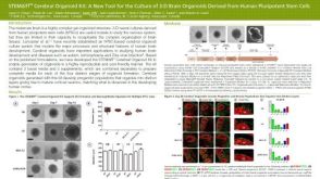 科学海报STEMdiff™ Cerebral Organoid Kit: A New Tool for the Culture of 3D Brain Organoids Derived from hPSCs
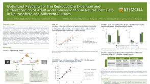


 沪公网安备31010102008431号