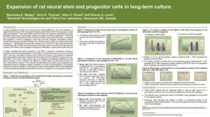
CGG-repeat dynamics and FMR1 gene silencing in fragile X syndrome stem cells and stem cell-derived neurons.
BACKGROUND Fragile X syndrome (FXS),a common cause of intellectual disability and autism,results from the expansion of a CGG-repeat tract in the 5' untranslated region of the FMR1 gene to<200 repeats. Such expanded alleles,known as full mutation (FM) alleles,are epigenetically silenced in differentiated cells thus resulting in the loss of FMRP,a protein important for learning and memory. The timing of repeat expansion and FMR1 gene silencing is controversial. METHODS We monitored the repeat size and methylation status of FMR1 alleles with expanded CGG repeats in patient-derived induced pluripotent stem cells (iPSCs) and embryonic stem cells (ESCs) that were grown for extended period of time either as stem cells or differentiated into neurons. We used a PCR assay optimized for the amplification of large CGG repeats for sizing,and a quantitative methylation-specific PCR for the analysis of FMR1 promoter methylation. The FMR1 mRNA levels were analyzed by qRT-PCR. FMRP levels were determined by western blotting and immunofluorescence. Chromatin immunoprecipitation was used to study the association of repressive histone marks with the FMR1 gene in FXS ESCs. RESULTS We show here that while FMR1 gene silencing can be seen in FXS embryonic stem cells (ESCs),some silenced alleles contract and when the repeat number drops below ˜400,DNA methylation erodes,even when the repeat number remains<200. The resultant active alleles do not show the large step-wise expansions seen in stem cells from other repeat expansion diseases. Furthermore,there may be selection against large active alleles and these alleles do not expand further or become silenced on neuronal differentiation. CONCLUSIONS Our data support the hypotheses that (i) large expansions occur prezygotically or in the very early embryo,(ii) large unmethylated alleles may be deleterious in stem cells,(iii) methylation can occur on alleles with<400 repeats very early in embryogenesis,and (iv) expansion and contraction may occur by different mechanisms. Our data also suggest that the threshold for stable methylation of FM alleles may be higher than previously thought. A higher threshold might explain why some carriers of FM alleles escape methylation. It may also provide a simple explanation for why silencing has not been observed in mouse models with<200 repeats.
View Publication
产品号#:
05832
05850
05857
05870
05875
85850
85857
85870
85875
产品名:
STEMdiff™ 神经花环选择试剂
mTeSR™1
mTeSR™1
Gundemir S et al. (SEP 2016)
Neuro-Oncology now157
The complex role of transglutaminase 2 in glioblastoma proliferation
BACKGROUND Glioblastomas (GBMs) are a heterogeneous group of primary brain tumors. These tumors are resistant to therapeutic interventions and invariably recur after surgical resection. The multifunctional protein transglutaminase 2 (TG2) has been shown to promote cell survival in a number of different tumors. There is also evidence that TG2 may be a pro-survival factor in GBMs. However,the roles that TG2 plays in facilitating GBM survival and proliferation have not yet been clearly delineated . METHODS The functions of TG2 are often cell- and context-specific. Therefore,in this study we examined the ability of TG2 to facilitate GBM proliferation using colony formation assays and 5-ethynyl-2'-deoxyuridine (EdU) incorporation in several different GBM cell lines as well as neurospheres derived from patient tumors representing the 3 major subtypes of GBM tumors (mesenchymal,proneural,and classical) and maintained in the absence of serum. TG2 knockdown or selective TG2 inhibitors were used to modulate TG2 expression and activity. RESULTS We show that TG2 plays differential roles in the proliferative process depending on the cell type. In most,but not all,GBM models TG2 plays a crucial role in the proliferative process,and some but not all TG2 inhibitors were highly effective at reducing proliferation in a large subset of the GBM models. CONCLUSION Our results show that TG2 plays an important-but notoriously context-specific-role in GBM cell biology. Nonetheless,as future studies unravel the genetic fingerprints" that make TG2 inhibitors effective this information could be exploited to develop TG2 inhibitors into personalized GBM therapies.
View Publication
产品号#:
05750
05751
产品名:
NeuroCult™ NS-A 基础培养基(人)
NeuroCult™ NS-A 扩增试剂盒(人)
Orr ME et al. (JUN 2012)
PLoS ONE 7 6 e39328
Genotype-Specific Differences between Mouse CNS Stem Cell Lines Expressing Frontotemporal Dementia Mutant or Wild Type Human Tau
Stem cell (SC) lines that capture the genetics of disease susceptibility provide new research tools. To assess the utility of mouse central nervous system (CNS) SC-containing neurosphere cultures for studying heritable neurodegenerative disease,we compared neurosphere cultures from transgenic mice that express human tau with the P301L familial frontotemporal dementia (FTD) mutation,rTg(tau(P301L))4510,with those expressing comparable levels of wild type human tau,rTg(tau(wt))21221. rTg(tau(P301L))4510 mice express the human tau(P301L) variant in their forebrains and display cellular,histological,biochemical and behavioral abnormalities similar to those in human FTD,including age-dependent differences in tau phosphorylation that distinguish them from rTg(tau(wt))21221 mice. We compared FTD-hallmark tau phosphorylation in neurospheres from rTg(tau(P301L))4510 mice and from rTg(tau(wt))21221 mice. The tau genotype-specific phosphorylation patterns in neurospheres mimicked those seen in mice,validating use of neurosphere cultures as models for studying tau phosphorylation. Genotype-specific tau phosphorylation was observed in 35 independent cell lines from individual fetuses; tau in rTg(tau(P301L))4510 cultures was hypophosphorylated in comparison with rTg(tau(wt))21221 as was seen in young adult mice. In addition,there were fewer human tau-expressing cells in rTg(tau(P301L))4510 than in rTg(tau(wt))21221 cultures. Following differentiation,neuronal filopodia-spine density was slightly greater in rTg(tau(P301L))4510 than rTg(tau(wt))21221 and control cultures. Together with the recapitulation of genotype-specific phosphorylation patterns,the observation that neurosphere lines maintained their cell line-specific-differences and retained SC characteristics over several passages supports the utility of SC cultures as surrogates for analysis of cellular disease mechanisms.
View Publication
产品号#:
05700
05701
05702
产品名:
NeuroCult™ 基础培养基(小鼠和大鼠)
NeuroCult™ 扩增添加物(小鼠和大鼠)
NeuroCult™扩增试剂盒(小鼠和大鼠)
Walker TL et al. (JAN 2011)
PloS one 6 3 e18153
The latent stem cell population is retained in the hippocampus of transgenic Huntington's disease mice but not wild-type mice.
The demonstration of the brain's ability to initiate repair in response to disease or injury has sparked considerable interest in therapeutic strategies to stimulate adult neurogenesis. In this study we examined the effect of a progressive neurodegenerative condition on neural precursor activity in the subventricular zone (SVZ) and hippocampus of the R6/1 transgenic mouse model of Huntington's disease (HD). Our results revealed an age-related decline in SVZ precursor numbers in both wild-type (WT) and HD mice. Interestingly,hippocampal precursor numbers declined with age in WT mice,although we observed maintenance in hippocampal precursor number in the HD animals in response to advancement of the disease. This maintenance was consistent with activation of a recently identified latent hippocampal precursor population. We found that the small latent stem cell population was also maintained in the HD hippocampus at 33 weeks,whereas it was not present in the WT. Our findings demonstrate that,despite a loss of neurogenesis in the HD hippocampus in vivo,there is a unique maintenance of the precursor and stem cells,which may potentially be activated to ameliorate disease symptoms.
View Publication
产品号#:
05700
05701
05702
产品名:
NeuroCult™ 基础培养基(小鼠和大鼠)
NeuroCult™ 扩增添加物(小鼠和大鼠)
NeuroCult™扩增试剂盒(小鼠和大鼠)
Poornima V et al. (MAR 2012)
Journal of molecular neuroscience : MN 46 3 585--94
P2X7 receptor-pannexin 1 hemichannel association: effect of extracellular calcium on membrane permeabilization.
Activation of P2X(7) receptor (P2X(7)R) and pannexin have been implicated in membrane permeabilization associated with ischemic cell death and many other inflammatory processes. P2X(7)R has a unique property of forming large pore upon repeated or prolonged application of agonist like ATP or 2',3'-(4-benzoyl) benzoyl ATP. It has been proposed that pannexin 1 (panx1) hemichannel associates with P2X(7)R to form large pore,though the actual mechanism is not yet understood. Calcium concentration in extracellular milieu drops in many patho-physiological conditions,e.g. ischemia,when P2X(7)R/pannexin is also known to be activated. Therefore,we hypothesize that extracellular calcium ([Ca(2+)](o)) plays an important role in the coupling of P2X(7)R-panx1 and subsequent membrane permeabilization. In this study we show that membrane permeability of the P2X(7)R and panx1 expressing N2A cell increases in ([Ca(2+)](o))-free solution. In [Ca(2+)](o)-free solution,fluorescent dye calcein trapped cells exhibited time-dependent dye leakage resulting in about 50% decrease of fluorescence intensity in 30 min. Control cells in 2 mM [Ca(2+)](o) did not show such leakage. Like N2A cells,mixed culture of neuron and glia,derived from hippocampal progenitor cells showed similar dye leakage. Dye leakage was blocked either by pannexin-specific blocker,carbenoxolone or P2X(7)R antagonists,Brilliant Blue G,and oxidized ATP. Furthermore P2X(7)R and panx1 were co-immunoprecipitated. The amount of P2X(7)R protein pulled-down with panx1,increased by twofold when cells were incubated 30 min in [Ca(2+)](o)-free buffer. Taken together,the results of this study demonstrate the activation and association of P2X(7)R-panx1,triggered by the removal of [Ca(2+)](o).
View Publication
产品号#:
05700
05701
05702
产品名:
NeuroCult™ 基础培养基(小鼠和大鼠)
NeuroCult™ 扩增添加物(小鼠和大鼠)
NeuroCult™扩增试剂盒(小鼠和大鼠)
Blackmore DG et al. (JAN 2012)
Scientific reports 2 250
Growth hormone responsive neural precursor cells reside within the adult mammalian brain.
The detection of growth hormone (GH) and its receptor in germinal regions of the mammalian brain prompted our investigation of GH and its role in the regulation of endogenous neural precursor cell activity. Here we report that the addition of exogenous GH significantly increased the expansion rate in long-term neurosphere cultures derived from wild-type mice,while neurospheres derived from GH null mice exhibited a reduced expansion rate. We also detected a doubling in the frequency of large (i.e. stem cell-derived) colonies for up to 120 days following a 7-day intracerebroventricular infusion of GH suggesting the activation of endogenous stem cells. Moreover,gamma irradiation induced the ablation of normally quiescent stem cells in GH-infused mice,resulting in a decline in olfactory bulb neurogenesis. These results suggest that GH activates populations of resident stem and progenitor cells,and therefore may represent a novel therapeutic target for age-related neurodegeneration and associated cognitive decline.
View Publication
产品号#:
05700
05701
05702
05740
产品名:
NeuroCult™ 基础培养基(小鼠和大鼠)
NeuroCult™ 扩增添加物(小鼠和大鼠)
NeuroCult™扩增试剂盒(小鼠和大鼠)
Ross HH et al. (MAY 2012)
Experimental neurology 235 1 238--45
In vivo intermittent hypoxia elicits enhanced expansion and neuronal differentiation in cultured neural progenitors.
In vitro exposure of neural progenitor cell (NPC) populations to reduced O(2) (e.g. 3% versus 20%) can increase their proliferation,survival and neuronal differentiation. Our objective was to determine if an acute (textless1hr),in vivo exposure to intermittent hypoxia (AIH) alters expansion and/or differentiation of subsequent in vitro cultures of NPC from the subventricular zone (SVZ). Neonatal C57BL/6 mice (postnatal day 4) were exposed to an AIH paradigm (20×1 minute; alternating 21% and 10% O(2)). Immediately after AIH,SVZ tissue was isolated and NPC populations were cultured and assayed either as neurospheres (NS) or as adherent monolayer cells (MASC). AIH markedly increased the capacity for expansion of cultured NS and MASC,and this was accompanied by increases in a proliferation maker (Ki67),MTT activity and hypoxia-inducible factor-1α (HIF-1α) signaling in NS cultures. Peptide blockade experiments confirmed that proteins downstream of HIF-1α are important for both proliferation and morphological changes associated with terminal differentiation in NS cultures. Finally,immunocytochemistry and Western blotting experiments demonstrated that AIH increased expression of the neuronal fate determination transcription factor Pax6 in SVZ tissue,and this was associated with increased neuronal differentiation in cultured NS and MASC. We conclude that in vivo AIH exposure can enhance the viability of subsequent in vitro SVZ-derived NPC cultures. AIH protocols may therefore provide a means to prime" NPC prior to transplantation into the injured central nervous system."
View Publication
产品号#:
05700
05701
05702
产品名:
NeuroCult™ 基础培养基(小鼠和大鼠)
NeuroCult™ 扩增添加物(小鼠和大鼠)
NeuroCult™扩增试剂盒(小鼠和大鼠)
Biasini E et al. (JAN 2012)
PloS one 7 3 e33472
The toxicity of a mutant prion protein is cell-autonomous, and can be suppressed by wild-type prion protein on adjacent cells.
Insight into the normal function of PrP(C),and how it can be subverted to produce neurotoxic effects,is provided by PrP molecules carrying deletions encompassing the conserved central region. The most neurotoxic of these mutants,Δ105-125 (called ΔCR),produces a spontaneous neurodegenerative illness when expressed in transgenic mice,and this phenotype can be dose-dependently suppressed by co-expression of wild-type PrP. Whether the toxic activity of ΔCR PrP and the protective activity or wild-type PrP are cell-autonomous,or can be exerted on neighboring cells,is unknown. To investigate this question,we have utilized co-cultures of differentiated neural stem cells derived from mice expressing ΔCR or wild-type PrP. Cells from the two kinds of mice,which are marked by the presence or absence of GFP,are differentiated together to yield neurons,astrocytes,and oligodendrocytes. As a surrogate read-out of ΔCR PrP toxicity,we assayed sensitivity of the cells to the cationic antibiotic,Zeocin. In a previous study,we reported that cells expressing ΔCR PrP are hypersensitive to the toxic effects of several cationic antibiotics,an effect that is suppressed by co-expression of wild type PrP,similar to the rescue of the neurodegenerative phenotype observed in transgenic mice. Using this system,we find that while ΔCR-dependent toxicity is cell-autonomous,the rescuing activity of wild-type PrP can be exerted in trans from nearby cells. These results provide important insights into how ΔCR PrP subverts a normal physiological function of PrP(C),and the cellular mechanisms underlying the rescuing process.
View Publication
产品号#:
05700
05701
05702
05703
05704
产品名:
NeuroCult™ 基础培养基(小鼠和大鼠)
NeuroCult™ 扩增添加物(小鼠和大鼠)
NeuroCult™扩增试剂盒(小鼠和大鼠)
NeuroCult™ 分化添加物(小鼠和大鼠)
NeuroCult™ 分化试剂盒(小鼠和大鼠)
Vukovic J et al. (MAY 2012)
The Journal of neuroscience : the official journal of the Society for Neuroscience 32 19 6435--43
Microglia modulate hippocampal neural precursor activity in response to exercise and aging.
Exercise has been shown to positively augment adult hippocampal neurogenesis; however,the cellular and molecular pathways mediating this effect remain largely unknown. Previous studies have suggested that microglia may have the ability to differentially instruct neurogenesis in the adult brain. Here,we used transgenic Csf1r-GFP mice to investigate whether hippocampal microglia directly influence the activation of neural precursor cells. Our results revealed that an exercise-induced increase in neural precursor cell activity was mediated via endogenous microglia and abolished when these cells were selectively removed from hippocampal cultures. Conversely,microglia from the hippocampi of animals that had exercised were able to activate latent neural precursor cells when added to neurosphere preparations from sedentary mice. We also investigated the role of CX(3)CL1,a chemokine that is known to provide a more neuroprotective microglial phenotype. Intraparenchymal infusion of a blocking antibody against the CX(3)CL1 receptor,CX(3)CR1,but not control IgG,dramatically reduced the neurosphere formation frequency in mice that had exercised. While an increase in soluble CX(3)CL1 was observed following running,reduced levels of this chemokine were found in the aged brain. Lower levels of CX(3)CL1 with advancing age correlated with the natural decline in neural precursor cell activity,a state that could be partially alleviated through removal of microglia. These findings provide the first direct evidence that endogenous microglia can exert a dual and opposing influence on neural precursor cell activity within the hippocampus,and that signaling through the CX(3)CL1-CX(3)CR1 axis critically contributes toward this process.
View Publication
Overexpression of calcium-permeable glutamate receptors in glioblastoma derived brain tumor initiating cells.
Glioblastoma multiforme is the most malignant type of primary brain tumor with a poor prognosis. These tumors consist of a heterogeneous population of malignant cells,including well-differentiated tumor cells and less differentiated cells with stem cell properties. These cancer stem cells,known as brain tumor initiating cells,likely contribute to glioma recurrence,as they are highly invasive,mobile,resistant to radiation and chemotherapy,and have the capacity to self-renew. Glioblastoma tumor cells release excitotoxic levels of glutamate,which may be a key process in the death of peritumoral neurons,formation of necrosis,local inflammation,and glioma-related seizures. Moreover,elevated glutamate levels in the tumor may act in paracrine and autocrine manner to activate glutamate receptors on glioblastoma tumor cells,resulting in proliferation and invasion. Using a previously described culturing condition that selectively promotes the growth of brain tumor initiating cells,which express the stem cell markers nestin and SOX-2,we characterize the expression of α-amino-3-hydroxy-5-methyl-4-isozolepropionic acid (AMPA)-type glutamate receptor subunits in brain tumor initiating cells derived from glioblastomas. Here we show for the first time that glioblastoma brain tumor initiating cells express high concentrations of functional calcium-permeable AMPA receptors,compared to the differentiated tumor cultures consisting of non-stem cells. Up-regulated calcium-permeable AMPA receptor expression was confirmed by immunoblotting,immunocytochemistry,and intracellular calcium imaging in response to specific agonists. Our findings raise the possibility that glutamate secretion in the GBM tumor microenvironment may stimulate brain tumor derived cancer stem cells.
View Publication

 EasySep™小鼠TIL(CD45)正选试剂盒


 沪公网安备31010102008431号