Erythropoietin (EPO) regulates the proliferation and differentiation of erythroid cells by binding to its specific transmembrane receptor (EPOR). The presence of EPO and its receptor in the CNS suggests a different function for EPO other than erythropoiesis. The purpose of the present study was to examine EPOR expression and the role of EPO in the proliferation of neonatal spinal cord-derived neural progenitor cells. The effect of EPO on cell cycle progression was also examined,as well as the signaling cascades involved in this process. Our results showed that EPOR was present in the neural progenitor cells and EPO significantly enhanced their proliferation. Cell cycle analysis of EPO-treated neural progenitor cells indicated a reduced percentage of cells in G0/G1 phase,whereas the cell proliferation index (S phase plus G2/M phase) was increased. EPO also increased the proportion of 5-bromo-2-deoxyuridine (BrdU)-positive cells. With respect to the cell cycle signaling,we examined the cyclin-dependent kinases D1,D2 and E,and cyclin-dependent kinase inhibitors,p21cip1,p27kip1 and p57kip2. No significant differences were observed in the expression of these transcripts after EPO administration. Interestingly,the anti-apoptotic factors,mcl-1 and bcl-2 were significantly increased twofold. Moreover,these specific effects of EPO were eliminated by incubation of the progenitor cells with anti-EPO neutralizing antibody. Those observations suggested that EPO may play a role in normal spinal cord development by regulating cell proliferation and apoptosis.
View Publication
产品号#:
05771
产品名:
Elliott E and Ginzburg I (JAN 2009)
FEBS letters 583 1 229--34
BAG-1 is preferentially expressed in neuronal precursor cells of the adult mouse brain and regulates their proliferation in vitro.
BAG-1 protein has been well characterized as necessary for proper neuronal development. However,little is known about the function of BAG-1 in the adult brain. In this work,the expression and localization of BAG-1 in the mature mouse brain was studied. The levels of both BAG-1 isoforms decrease significantly in the brain during development. BAG-1 was found preferentially expressed in Neuronal Precursor Cells (NPCs) in the two major niches of neurogenesis. Lentiviral mediated overexpression of BAG-1 increased the proliferation rate of cultured NPCs. In addition,depletion of BAG-1 from NPCs induced a decrease in NPCs proliferation in the presence of a stress hormone,corticosterone. These data suggest a role for BAG-1 in mechanisms of neurogenesis in the adult mouse brain.
View Publication
产品号#:
05700
05701
05702
产品名:
NeuroCult™ 基础培养基(小鼠和大鼠)
NeuroCult™ 扩增添加物(小鼠和大鼠)
NeuroCult™扩增试剂盒(小鼠和大鼠)
Yan Y et al. (FEB 2015)
1341 257--284
Generation of Neural Progenitor Spheres from Human Pluripotent Stem Cells in a Suspension Bioreactor
Conventional two-dimensional (2-D) culture systems cannot provide large numbers of human pluripotent stem cells (hPSCs) and their derivatives that are demanded for commercial and clinical applications in in vitro drug screening,disease modeling,and potentially cell therapy. The technologies that support three-dimensional (3-D) suspension culture,such as a stirred bioreactor,are generally considered as promising approaches to produce the required cells. Recently,suspension bioreactors have also been used to generate mini-brain-like structure from hPSCs for disease modeling,showing the important role of bioreactor in stem cell culture. This chapter describes a detailed culture protocol for neural commitment of hPSCs into neural progenitor cell (NPC) spheres using a spinner bioreactor. The basic steps to prepare hPSCs for bioreactor inoculation are illustrated from cell thawing to cell propagation. The method for generating NPCs from hPSCs in the spinner bioreactor along with the static control is then described. The protocol in this study can be applied to the generation of NPCs from hPSCs for further neural subtype specification,3-D neural tissue development,or potential preclinical studies or clinical applications in neurological diseases.
View Publication
产品号#:
05850
05857
05870
05875
72302
72304
72307
72308
85850
85857
85870
85875
100-1044
产品名:
Y-27632(二盐酸盐)
Y-27632(二盐酸盐)
Y-27632(二盐酸盐)
Y-27632(二盐酸盐)
mTeSR™1
mTeSR™1
Y-27632(二盐酸盐)
Miranda C et al. (OCT 2015)
Biotechnology Journal 10 10 1612--1624
Spatial and temporal control of cell aggregation efficiently directs human pluripotent stem cells towards neural commitment
3D suspension culture is generally considered a promising method to achieve efficient expansion and controlled differentiation of human pluripotent stem cells (hPSCs). In this work,we focused on developing an integrated culture platform for expansion and neural commitment of hPSCs into neural precursors using 3D suspension conditions and chemically-defined culture media. We evaluated different inoculation methodologies for hPSC expansion as 3D aggregates and characterized the resulting cultures in terms of aggregate size distribution. It was demonstrated that upon single-cell inoculation,after four days of culture,3D aggregates were composed of homogenous populations of hPSC and were characterized by an average diameter of 139 ± 26 μm,which was determined to be the optimal size to initiate neural commitment. Temporal analysis revealed that upon neural specification it is possible to maximize the percentage of neural precursor cells expressing the neural markers Sox1 and Pax6 after nine days of culture. These results highlight our ability to define a robust method for production of hPSC-derived neural precursors that minimizes processing steps and that constitutes a promising alternative to the traditional planar adherent culture system due to a high potential for scaling-up.
View Publication
产品号#:
05850
05857
05870
05875
27305
85850
85857
85870
85875
产品名:
mTeSR™1
mTeSR™1
Gallegos-Cá et al. (AUG 2015)
Stem cells and development 24 16 1901--1911
For diseases of the brain,the pig (Sus scrofa) is increasingly being used as a model organism that shares many anatomical and biological similarities with humans. We report that pig induced pluripotent stem cells (iPSC) can recapitulate events in early mammalian neural development. Pig iPSC line (POU5F1(high)/SSEA4(low)) had a higher potential to form neural rosettes (NR) containing neuroepithelial cells than either POU5F1(low)/SSEA4(low) or POU5F1(low)/SSEA4(high) lines. Thus,POU5F1 and SSEA4 pluripotency marker profiles in starting porcine iPSC populations can predict their propensity to form more robust NR populations in culture. The NR were isolated and expanded in vitro,retaining their NR morphology and neuroepithelial molecular properties. These cells expressed anterior central nervous system fate markers OTX2 and GBX2 through at least seven passages,and responded to retinoic acid,promoting a more posterior fate (HOXB4+,OTX2-,and GBX2-). These findings offer insight into pig iPSC development,which parallels the human iPSC in both anterior and posterior neural cell fates. These in vitro similarities in early neural differentiation processes support the use of pig iPSC and differentiated neural cells as a cell therapy in allogeneic porcine neural injury and degeneration models,providing relevant translational data for eventual human neural cell therapies.
View Publication
产品号#:
05850
05857
05870
05875
07923
85850
85857
85870
85875
产品名:
Dispase (1 U/mL)
mTeSR™1
mTeSR™1
Su CTE et al. (FEB 2015)
Journal of visualized experiments : JoVE 96 1--9
An Optogenetic Approach for Assessing Formation of Neuronal Connections in a Co-culture System.
Here we describe a protocol to generate a co-culture consisting of 2 different neuronal populations. Induced pluripotent stem cells (iPSCs) are reprogrammed from human fibroblasts using episomal vectors. Colonies of iPSCs can be observed 30 days after initiation of fibroblast reprogramming. Pluripotent colonies are manually picked and grown in neural induction medium to permit differentiation into neural progenitor cells (NPCs). iPSCs rapidly convert into neuroepithelial cells within 1 week and retain the capability to self-renew when maintained at a high culture density. Primary mouse NPCs are differentiated into astrocytes by exposure to a serum-containing medium for 7 days and form a monolayer upon which embryonic day 18 (E18) rat cortical neurons (transfected with channelrhodopsin-2 (ChR2)) are added. Human NPCs tagged with the fluorescent protein,tandem dimer Tomato (tdTomato),are then seeded onto the astrocyte/cortical neuron culture the following day and allowed to differentiate for 28 to 35 days. We demonstrate that this system forms synaptic connections between iPSC-derived neurons and cortical neurons,evident from an increase in the frequency of synaptic currents upon photostimulation of the cortical neurons. This co-culture system provides a novel platform for evaluating the ability of iPSC-derived neurons to create synaptic connections with other neuronal populations.
View Publication
Paulsen BdS et al. (APR 2014)
Schizophrenia Research 154 1-3 30--35
Valproate reverts zinc and potassium imbalance in schizophrenia-derived reprogrammed cells
Schizophrenia has been considered a devastating clinical syndrome rather than a single disease. Nevertheless,the mechanisms behind the onset of schizophrenia have been only partially elucidated. Several studies propose that levels of trace elements are abnormal in schizophrenia; however,conflicting data generated from different biological sources prevent conclusions being drawn. In this work,we used synchrotron radiation X-ray microfluorescence spectroscopy to compare trace element levels in neural progenitor cells (NPCs) derived from two clones of induced pluripotent stem cell lines of a clozapine-resistant schizophrenic patient and two controls. Our data reveal the presence of elevated levels of potassium and zinc in schizophrenic NPCs. Neural cells treated with valproate,an adjunctive medication for schizophrenia,brought potassium and zinc content back to control levels. These results expand the understanding of atomic element imbalance related to schizophrenia and may provide novel insights for the screening of drugs to treat mental disorders. ?? 2014 Elsevier B.V.
View Publication
产品号#:
05850
05857
05870
05875
85850
85857
85870
85875
产品名:
mTeSR™1
mTeSR™1
Binder LI et al. (SEP 1984)
Proceedings of the National Academy of Sciences of the United States of America 81 17 5613--7
Heterogeneity of microtubule-associated protein 2 during rat brain development.
The electrophoretic pattern of the large microtubule-associated protein,MAP2,changes during rat brain development. Immunoblots of NaDodSO4 extracts obtained from the cerebral cortex,cerebellum,and thalamus at 10-15 days after birth reveal only a single electrophoretic species when probed with any of three MAP2 monoclonal antibodies. By contrast,adult MAP2 contains two immunoreactive species,MAP2a and MAP2b. The single band of MAP2 from immature brain electrophoretically comigrates with adult MAP2b. Between postnatal days 17 and 18,immature MAP2 simultaneously resolves into two species in both the cerebellum and cerebral cortex. Immunoblots of NaDodSO4 extracts from spinal cord demonstrate the adult complement of MAP2 by day 10,indicating that MAP2 does not change coordinately throughout the entire central nervous system. In vitro cAMP-dependent phosphorylation of immature MAP2 causes a band split reminiscent of that seen during brain development in vivo. The possibility that the developmentally regulated changes observed in MAP2 during brain maturation are due to timed phosphorylation events is discussed.
View Publication
产品号#:
01410
产品名:
Lippmann ES et al. (APR 2014)
Stem Cells 32 4 1032--1042
Defined human pluripotent stem cell culture enables highly efficient neuroepithelium derivation without small molecule inhibitors.
The embryonic neuroepithelium gives rise to the entire central nervous system in vivo,making it an important tissue for developmental studies and a prospective cell source for regenerative applications. Current protocols for deriving homogenous neuroepithelial cultures from human pluripotent stem cells (hPSCs) consist of either embryoid body-mediated neuralization followed by a manual isolation step or adherent differentiation using small molecule inhibitors. Here,we report that hPSCs maintained under chemically defined,feeder-independent,and xeno-free conditions can be directly differentiated into pure neuroepithelial cultures ([mt]90% Pax6(+)/N-cadherin(+) with widespread rosette formation) within 6 days under adherent conditions,without small molecule inhibitors,and using only minimalistic medium consisting of Dulbecco's modified Eagle's medium/F-12,sodium bicarbonate,selenium,ascorbic acid,transferrin,and insulin (i.e.,E6 medium). Furthermore,we provide evidence that the defined culture conditions enable this high level of neural conversion in contrast to hPSCs maintained on mouse embryonic fibroblasts (MEFs). In addition,hPSCs previously maintained on MEFs could be rapidly converted to a neural compliant state upon transfer to these defined conditions while still maintaining their ability to generate all three germ layers. Overall,this fully defined and scalable protocol should be broadly useful for generating therapeutic neural cells for regenerative applications.
View Publication
产品号#:
05850
05857
05870
05875
85850
85857
85870
85875
产品名:
mTeSR™1
mTeSR™1
Qu Q et al. (MAR 2014)
Nature communications 5 3449
High-efficiency motor neuron differentiation from human pluripotent stem cells and the function of Islet-1.
Efficient derivation of large-scale motor neurons (MNs) from human pluripotent stem cells is central to the understanding of MN development,modelling of MN disorders in vitro and development of cell-replacement therapies. Here we develop a method for rapid (20 days) and highly efficient (˜70%) differentiation of mature and functional MNs from human pluripotent stem cells by tightly modulating neural patterning temporally at a previously undefined primitive neural progenitor stage. This method also allows high-yield (textgreater250%) MN production in chemically defined adherent cultures. Furthermore,we show that Islet-1 is essential for formation of mature and functional human MNs,but,unlike its mouse counterpart,does not regulate cell survival or suppress the V2a interneuron fate. Together,our discoveries improve the strategy for MN derivation,advance our understanding of human neural specification and MN development,and provide invaluable tools for human developmental studies,drug discovery and regenerative medicine.
View Publication
产品号#:
05850
05857
05870
05875
85850
85857
85870
85875
产品名:
mTeSR™1
mTeSR™1
Hotta R et al. (APR 2016)
Neurogastroenterology and motility : the official journal of the European Gastrointestinal Motility Society 28 4 498--512
Isogenic enteric neural progenitor cells can replace missing neurons and glia in mice with Hirschsprung disease.
BACKGROUND Transplanting autologous patient-derived enteric neuronal stem/progenitor cells (ENSCs) is an innovative approach to replacing missing enteric neurons in patients with Hirschsprung disease (HSCR). Using autologous cells eliminates immunologic and ethical concerns raised by other cell sources. However,whether postnatal aganglionic bowel is permissive for transplanted ENSCs and whether ENSCs from HSCR patients can be successfully isolated,cultured,and transplanted in vivo remains unknown. METHODS ENSCs isolated from the ganglionic intestine of Ednrb(-/-) mice (HSCR-ENSCs) were characterized immunohistochemically and evaluated for their capacity to proliferate and differentiate in vitro. Fluorescently labeled ENSCs were co-cultured ex vivo with aganglionic Ednrb(-/-) colon. For in vivo transplantation,HSCR-ENSCs were labeled with lentivirus expressing green fluorescent protein (GFP) and implanted into aganglionic embryonic chick gut in ovo and postnatal aganglionic Ednrb(-/-) rectum in vivo. KEY RESULTS HSCR-ENSCs maintain normal capacity self-renewal and neuronal differentiation. Moreover,the Ednrb(-/-) aganglionic environment is permissive to engraftment by wild-type ENSCs ex vivo and supports migratrion and neuroglial differentiation of these cells following transplantation in vivo. Lentiviral GFP-labeled HSCR-ENSCs populated embryonic chick hindgut and postnatal colon of Ednrb(-/-) HSCR,with cells populating the intermuscular layer and forming enteric neurons and glia. CONCLUSIONS & INFERENCES ENSCs can be isolated and cultured from mice with HSCR,and transplanted into the aganglionic bowel of HSCR littermates to generate enteric neuronal networks. These results in an isogenic model establish the potential of using autologous-derived stem cells to treat HSCR and other intestinal neuropathies.
View Publication

 EasySep™小鼠TIL(CD45)正选试剂盒


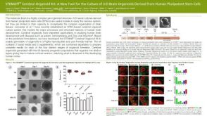 科学海报STEMdiff™ Cerebral Organoid Kit: A New Tool for the Culture of 3D Brain Organoids Derived from hPSCs
 沪公网安备31010102008431号