Human immunodeficiency virus-driven expansion of CD4+CD25+ regulatory T cells, which suppress HIV-specific CD4 T-cell responses in HIV-infected patients.
The present study demonstrates that CD4(+)CD25(+) T cells,expanded in peripheral blood of HIV-infected patients receiving highly active antiretroviral therapy (HAART),exhibit phenotypic,molecular,and functional characteristics of regulatory T cells. The majority of peripheral CD4(+)CD25(+) T cells from HIV-infected patients expressed a memory phenotype. They were found to constitutively express transcription factor forkhead box P3 (Foxp3) messengers. CD4(+)CD25(+) T cells weakly proliferated to immobilized anti-CD3 monoclonal antibody (mAb) and addition of soluble anti-CD28 mAb significantly increased proliferation. In contrast to CD4(+)CD25(-) T cells,CD4(+)CD25(+) T cells from HIV-infected patients did not proliferate in response to recall antigens and to p24 protein. The proliferative capacity of CD4 T cells to tuberculin,cytomegalovirus (CMV),and p24 significantly increased following depletion of CD4(+)CD25(+) T cells. Furthermore,addition of increasing numbers of CD4(+)CD25(+) T cells resulted in a dose-dependent inhibition of CD4(+)CD25(-) T-cell proliferation to tuberculin and p24. CD4(+)CD25(+) T cells responded specifically to p24 antigen stimulation by expressing transforming growth factor beta (TGF-beta) and interleukin 10 (IL-10),thus indicating the presence of p24-specific CD4(+) T cells among the CD4(+)CD25(+) T-cell subset. Suppressive activity was not dependent on the secretion of TGF-beta or IL-10. Taken together,our results suggest that persistence of HIV antigens might trigger the expansion of CD4(+)CD25(+) regulatory T cells,which might induce a tolerance to HIV in vivo.
View Publication
产品号#:
15022
15062
产品名:
RosetteSep™人CD4+ T细胞富集抗体混合物
RosetteSep™人CD4+ T细胞富集抗体混合物
Abdelwahab SF et al. (DEC 2003)
Proceedings of the National Academy of Sciences of the United States of America 100 25 15006--10
HIV-1-suppressive factors are secreted by CD4+ T cells during primary immune responses.
CD4+ T cells are required for immunity against many viral infections,including HIV-1 where a positive correlation has been observed between strong recall responses and low HIV-1 viral loads. Some HIV-1-specific CD4+ T cells are preferentially infected with HIV-1,whereas others escape infection by unknown mechanisms. One possibility is that some CD4+ T cells are protected from infection by the secretion of soluble HIV-suppressive factors,although it is not known whether these factors are produced during primary antigen-specific responses. Here,we show that soluble suppressive factors are produced against CXCR4 and CCR5 isolates of HIV-1 during the primary immune response of human CD4+ T cells. This activity requires antigenic stimulation of naïve CD4+ T cells. One anti-CXCR4 factor is macrophage-derived chemokine (chemokine ligand 22,CCL22),and anti-CCR5 factors include macrophage inflammatory protein-1 alpha (CCL3),macrophage inflammatory protein-1 beta (CCL4),and RANTES (regulated upon activation of normal T cells expressed and secreted) (CCL5). Intracellular staining confirms that CD3+CD4+ T cells are the source of the prototype HIV-1-inhibiting chemokines CCL22 and CCL4. These results show that CD4+ T cells secrete an evolving HIV-1-suppressive activity during the primary immune response and that this activity is comprised primarily of CC chemokines. The data also suggest that production of such factors should be considered in the design of vaccines against HIV-1 and as a mechanism whereby the host can control infections with this virus.
View Publication
产品号#:
09500
09600
09650
19155
19155RF
产品名:
BIT 9500血清替代物
StemSpan™ SFEM
StemSpan™ SFEM
Jones DC et al. (JUL 2003)
Journal of immunology 171 1 196--203
Peroxisome proliferator-activated receptor alpha negatively regulates T-bet transcription through suppression of p38 mitogen-activated protein kinase activation.
Expression of the nuclear hormone receptor peroxisome proliferator-activated receptor alpha (PPARalpha) in resting lymphocytes was recently established,although the physiologic role(s) played by this nuclear hormone receptor in these cell types remains unresolved. In this study,we used CD4(+) T cells isolated from PPARalpha(-/-) and wild-type mice,as well as cell lines that constitutively express PPARalpha,in experiments designed to evaluate the role of this hormone receptor in the regulation of T cell function. We report that activated CD4(+) T cells lacking PPARalpha produce increased levels of IFN-gamma,but significantly lower levels of IL-2 when compared with activated wild-type CD4(+) T cells. Furthermore,we demonstrate that PPARalpha regulates the expression of these cytokines by CD4(+) T cells in part,through its ability to negatively regulate the transcription of T-bet. The induction of T-bet expression in CD4(+) T cells was determined to be positively influenced by p38 mitogen-activated protein (MAP) kinase activation,and the presence of unliganded PPARalpha effectively suppressed the phosphorylation of p38 MAP kinase. The activation of PPARalpha with highly specific ligands relaxed its capacity to suppress p38 MAP kinase phosphorylation and promoted T-bet expression. These results demonstrate a novel DNA-binding independent and agonist-controlled regulatory influence by the nuclear hormone receptor PPARalpha.
View Publication
产品号#:
03814
产品名:
ClonaCell™-TCS培养基
Schlecht G et al. (OCT 2001)
Journal of immunology (Baltimore,Md. : 1950) 167 8 4215--21
Induction of CTL and nonpolarized Th cell responses by CD8alpha(+) and CD8alpha(-) dendritic cells.
Two distinct dendritic cell (DC) subpopulations have been evidenced in mice on the basis of their differential CD8alpha expression and their localization in lymphoid organs. Several reports suggest that CD8alpha(+) and CD8alpha(-) DC subsets could be functionally different. In this study,using a panel of MHC class I- and/or class II-restricted peptides,we analyzed CD4(+) and CD8(+) T cell responses obtained after i.v. injection of freshly purified peptide-pulsed DC subsets. First,we showed that both DC subsets efficiently induce specific CTL responses and Th1 cytokine production in the absence of CD4(+) T cell priming. Second,we showed that in vivo activation of CD4(+) T cells by CD8alpha(+) or CD8alpha(-) DC,injected i.v.,leads to a nonpolarized Th response with production of both Th1 and Th2 cytokines. The CD8alpha(-) subset induced a higher production of Th2 cytokines such as IL-4 and IL-10 than the CD8alpha(+) subset. However,IL-5 was produced by CD4(+) T cells activated by both DC subsets. When both CD4(+) and CD8(+) T cells were primed by DC injected i.v.,a similar pattern of cytokines was observed,but,under these conditions,Th1 cytokines were mainly produced by CD8(+) T cells,while Th2 cytokines were produced by CD4(+) T cells. Thus,this study clearly shows that CD4(+) T cell responses do not influence the development of specific CD8(+) T cell cytotoxic responses induced either by CD8alpha(+) or CD8alpha(-) DC subsets.
View Publication

 EasySep™小鼠TIL(CD45)正选试剂盒


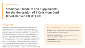 技术公告StemSpan™ Medium and Supplements for the Generation of T Cells from Cord Blood-Derived CD34+ Cells
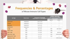 挂图Frequencies and Percentages of Mouse Immune Cell Types List of the frequencies of over 25 immune cell types in C57BL/6 mice
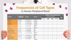 挂图血液相关来源中人细胞类型的比例 List of the frequencies of over 35 cell types in normal human blood-related sources.


 沪公网安备31010102008431号