Wang J et al. (NOV 2013)
Biomaterials 34 35 8878--8886
Effect of engineered anisotropy on the susceptibility of human pluripotent stem cell-derived ventricular cardiomyocytes to arrhythmias
Human (h) pluripotent stem cells (PSC) such as embryonic stem cells (ESC) can be directed into cardiomyocytes (CMs),representing a potential unlimited cell source for disease modeling,cardiotoxicity screening and myocardial repair. Although the electrophysiology of single hESC-CMs is now better defined,their multi-cellular arrhythmogenicity has not been thoroughly assessed due to the lack of a suitable experimental platform. Indeed,the generation of ventricular (V) fibrillation requires single-cell triggers as well as sustained multi-cellular reentrant events. Although native VCMs are aligned in a highly organized fashion such that electrical conduction is anisotropic for coordinated contractions,hESC-derived CM (hESC-CM) clusters are heterogenous and randomly organized,and therefore not representative of native conditions. Here,we reported that engineered alignment of hESC-VCMs on biomimetic grooves uniquely led to physiologically relevant responses. Aligned but not isotropic control preparations showed distinct longitudinal (L) and transverse (T) conduction velocities (CV),resembling the native human V anisotropic ratio (AR=LCV/TCV=1.8-2.0). Importantly,the total incidence of spontaneous and inducible arrhythmias significantly reduced from 57% in controls to 17-23% of aligned preparations,thereby providing a physiological baseline for assessing arrhythmogenicity. As such,promotion of pro-arrhythmic effect (e.g.,spatial dispersion by ?? adrenergic stimulation) could be better predicted. Mechanistically,such anisotropy-induced electrical stability was not due to maturation of the cellular properties of hESC-VCMs but their physical arrangement. In conclusion,not only do functional anisotropic hESC-VCMs engineered by multi-scale topography represent a more accurate model for efficacious drug discovery and development as well as arrhythmogenicity screening (of pharmacological and genetic factors),but our approach may also lead to future transplantable prototypes with improved efficacy and safety against arrhythmias. ?? 2013.
View Publication
产品号#:
05850
05857
05870
05875
85850
85857
85870
85875
产品名:
mTeSR™1
mTeSR™1
Haraguchi Y et al. (DEC 2015)
Journal of Tissue Engineering and Regenerative Medicine 9 12 1363--1375
Simple suspension culture system of human iPS cells maintaining their pluripotency for cardiac cell sheet engineering.
In this study,a simple three-dimensional (3D) suspension culture method for the expansion and cardiac differentiation of human induced pluripotent stem cells (hiPSCs) is reported. The culture methods were easily adapted from two-dimensional (2D) to 3D culture without any additional manipulations. When hiPSCs were directly applied to 3D culture from 2D in a single-cell suspension,only a few aggregated cells were observed. However,after 3 days,culture of the small hiPSC aggregates in a spinner flask at the optimal agitation rate created aggregates which were capable of cell passages from the single-cell suspension. Cell numbers increased to approximately 10-fold after 12 days of culture. The undifferentiated state of expanded hiPSCs was confirmed by flow cytometry,immunocytochemistry and quantitative RT-PCR,and the hiPSCs differentiated into three germ layers. When the hiPSCs were subsequently cultured in a flask using cardiac differentiation medium,expression of cardiac cell-specific genes and beating cardiomyocytes were observed. Furthermore,the culture of hiPSCs on Matrigel-coated dishes with serum-free medium containing activin A,BMP4 and FGF-2 enabled it to generate robust spontaneous beating cardiomyocytes and these cells expressed several cardiac cell-related genes,including HCN4,MLC-2a and MLC-2v. This suggests that the expanded hiPSCs might maintain the potential to differentiate into several types of cardiomyocytes,including pacemakers. Moreover,when cardiac cell sheets were fabricated using differentiated cardiomyocytes,they beat spontaneously and synchronously,indicating electrically communicative tissue. This simple culture system might enable the generation of sufficient amounts of beating cardiomyocytes for use in cardiac regenerative medicine and tissue engineering.
View Publication

 EasySep™小鼠TIL(CD45)正选试剂盒


 实验方案Optimizing Delivery Efficiency with Fluorescent Dextran Using the CellPore™ Transfection System
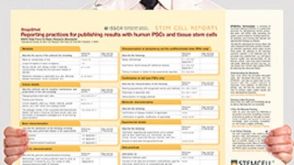 挂图Reporting Practices for Publishing Results with hPSCs Learn how to plan and conduct your human pluripotent stem cell (hPSC)-based research following the ISSCR’s Standards for Human Stem Cell Use in Research


 沪公网安备31010102008431号