Intrinsic Immunity Shapes Viral Resistance of Stem Cells.
Stem cells are highly resistant to viral infection compared to their differentiated progeny; however,the mechanism is mysterious. Here,we analyzed gene expression in mammalian stem cells and cells at various stages of differentiation. We find that,conserved across species,stem cells express a subset of genes previously classified as interferon (IFN) stimulated genes (ISGs) but that expression is intrinsic,as stem cells are refractory to interferon. This intrinsic ISG expression varies in a cell-type-specific manner,and many ISGs decrease upon differentiation,at which time cells become IFN responsive,allowing induction of a broad spectrum of ISGs by IFN signaling. Importantly,we show that intrinsically expressed ISGs protect stem cells against viral infection. We demonstrate the in vivo importance of intrinsic ISG expression for protecting stem cells and their differentiation potential during viral infection. These findings have intriguing implications for understanding stem cell biology and the evolution of pathogen resistance.
View Publication
产品号#:
02691
04434
04444
05110
05711
05712
05872
05873
18056
18056RF
72052
72054
72302
72304
72307
72308
70039
70039.1
70039.2
70039.3
70039.4
70039.5
70039.6
60062
60062BT
60062FI
60062FI.1
60062PE
60062PE.1
60045
60045AZ
60045AZ.1
60045BT
60045FI
60045FI.1
600
产品名:
StemSpan™ CD34+扩增添加物 (10X)
MethoCult™ H4434 Classic
MethoCult™ H4434 Classic
STEMdiff™定型内胚层检测试剂盒
NeuroCult™ SM1 神经添加物
CHIR99021
CHIR99021
Y-27632(二盐酸盐)
Y-27632(二盐酸盐)
Y-27632(二盐酸盐)
Y-27632(二盐酸盐)
抗人SSEA-4抗体,克隆号MC-813-70,生物素
抗人SSEA-4抗体,克隆号MC-813-70,FITC
抗人SSEA-4抗体, 克隆号MC-813-70,FITC
抗人SSEA-4抗体,克隆号MC-813-70,PE
抗人SSEA-4抗体,克隆号MC-813-70,PE
抗人CD90抗体,克隆5E10
抗人CD90抗体,克隆5E10,APC
抗人CD90抗体,克隆5E10,APC
抗人CD90抗体,克隆5E10,Biotin
抗人CD90抗体,克隆5E10,FITC
抗人CD90抗体,克隆5E10,FITC
Mariotti J et al. (JAN 2008)
Journal of immunology (Baltimore,Md. : 1950) 180 1 89--105
Ex vivo rapamycin generates apoptosis-resistant donor Th2 cells that persist in vivo and prevent hemopoietic stem cell graft rejection.
Because ex vivo rapamycin generates murine Th2 cells that prevent Graft-versus-host disease more potently than control Th2 cells,we hypothesized that rapamycin would generate Th2/Tc2 cells (Th2/Tc2.R cells) that abrogate fully MHC-disparate hemopoietic stem cell rejection more effectively than control Th2/Tc2 cells. In a B6-into-BALB/c graft rejection model,donor Th2/Tc2.R cells were indeed enriched in their capacity to prevent rejection; importantly,highly purified CD4+ Th2.R cells were also highly efficacious for preventing rejection. Rapamycin-generated Th2/Tc2 cells were less likely to die after adoptive transfer,accumulated in vivo at advanced proliferative cycles,and were present in 10-fold higher numbers than control Th2/Tc2 cells. Th2.R cells had a multifaceted,apoptosis-resistant phenotype,including: 1) reduced apoptosis after staurosporine addition,serum starvation,or CD3/CD28 costimulation; 2) reduced activation of caspases 3 and 9; and 3) increased anti-apoptotic Bcl-xL expression and reduced proapoptotic Bim and Bid expression. Using host-versus-graft reactivity as an immune correlate of graft rejection,we found that the in vivo efficacy of Th2/Tc2.R cells 1) did not require Th2/Tc2.R cell expression of IL-4,IL-10,perforin,or Fas ligand; 2) could not be reversed by IL-2,IL-7,or IL-15 posttransplant therapy; and 3) was intact after therapy with Th2.R cells relatively devoid of Foxp3 expression. We conclude that ex vivo rapamycin generates Th2 cells that are resistant to apoptosis,persist in vivo,and effectively prevent rejection by a mechanism that may be distinct from previously described graft-facilitating T cells.
View Publication
产品号#:
09600
09650
产品名:
StemSpan™ SFEM
StemSpan™ SFEM
Crist SA et al. (APR 2008)
Blood 111 7 3553--61
Nuclear factor of activated T cells (NFAT) mediates CD154 expression in megakaryocytes.
Platelets are an abundant source of CD40 ligand (CD154),an immunomodulatory and proinflammatory molecule implicated in the onset and progression of several inflammatory diseases,including systemic lupus erythematosus (SLE),diabetes,and cardiovascular disease. Heretofore considered largely restricted to activated T cells,we initiated studies to investigate the source and regulation of platelet-associated CD154. We found that CD154 is abundantly expressed in platelet precursor cells,megakaryocytes. We show that CD154 is expressed in primary human CD34+ and murine hematopoietic precursor cells only after cytokine-driven megakaryocyte differentiation. Furthermore,using several established megakaryocyte-like cells lines,we performed promoter analysis of the CD154 gene and found that NFAT,a calcium-dependent transcriptional regulator associated with activated T cells,mediated both differentiation-dependent and inducible megakaryocyte-specific CD154 expression. Overall,these data represent the first investigation of the regulation of a novel source of CD154 and suggests that platelet-associated CD154 can be biochemically modulated.
View Publication
产品号#:
09600
09650
产品名:
StemSpan™ SFEM
StemSpan™ SFEM
Schü et al. (MAY 2008)
Blood 111 9 4532--41
The MADS transcription factor Mef2c is a pivotal modulator of myeloid cell fate.
Mef2c is a MADS (MCM1-agamous-deficient serum response factor) transcription factor best known for its role in muscle and cardiovascular development. A causal role of up-regulated MEF2C expression in myelomonocytic acute myeloid leukemia (AML) has recently been demonstrated. Due to the pronounced monocytic component observed in Mef2c-induced AML,this study was designed to assess the importance of Mef2c in normal myeloid differentiation. Analysis of bone marrow (BM) cells manipulated to constitutively express Mef2c demonstrated increased monopoiesis at the expense of granulopoiesis,whereas BM isolated from Mef2c(Delta/-) mice showed reduced levels of monocytic differentiation in response to cytokines. Mechanistic studies showed that loss of Mef2c expression correlated with reduced levels of transcripts encoding c-Jun,but not PU.1,C/EBPalpha,or JunB transcription factors. Inhibiting Jun expression by short-interfering RNA impaired Mef2c-mediated inhibition of granulocyte development. Moreover,retroviral expression of c-Jun in BM cells promoted monocytic differentiation. The ability of Mef2c to modulate cell-fate decisions between monocyte and granulocyte differentiation,coupled with its functional sensitivity to extracellular stimuli,demonstrate an important role in immunity--and,consistent with findings of other myeloid transcription factors,a target of oncogenic lesions in AML.
View Publication
产品号#:
03434
03444
09600
09650
18556
18556RF
产品名:
MethoCult™ GF M3434
MethoCult™ GF M3434
StemSpan™ SFEM
StemSpan™ SFEM
Mirabelli P et al. (JAN 2008)
BMC physiology 8 1 13
Extended flow cytometry characterization of normal bone marrow progenitor cells by simultaneous detection of aldehyde dehydrogenase and early hematopoietic antigens: implication for erythroid differentiation studies.
BACKGROUND: Aldehyde dehydrogenase (ALDH) is a cytosolic enzyme highly expressed in hematopoietic precursors from cord blood and granulocyte-colony stimulating factor mobilized peripheral blood,as well as in bone marrow from patients with acute myeloblastic leukemia. As regards human normal bone marrow,detailed characterization of ALDH+ cells has been addressed by one single study (Gentry et al,2007). The goal of our work was to provide new information about the dissection of normal bone marrow progenitor cells based upon the simultaneous detection by flow cytometry of ALDH and early hematopoietic antigens,with particular attention to the expression of ALDH on erythroid precursors. To this aim,we used three kinds of approach: i) multidimensional analytical flow cytometry,detecting ALDH and early hematopoietic antigens in normal bone marrow; ii) fluorescence activated cell sorting of distinct subpopulations of progenitor cells,followed by in vitro induction of erythroid differentiation; iii) detection of ALDH+ cellular subsets in bone marrow from pure red cell aplasia patients. RESULTS: In normal bone marrow,we identified three populations of cells,namely ALDH+CD34+,ALDH-CD34+ and ALDH+CD34- (median percentages were 0.52,0.53 and 0.57,respectively). As compared to ALDH-CD34+ cells,ALDH+CD34+ cells expressed the phenotypic profile of primitive hematopoietic progenitor cells,with brighter expression of CD117 and CD133,accompanied by lower display of CD38 and CD45RA. Of interest,ALDH+CD34- population disclosed a straightforward erythroid commitment,on the basis of three orders of evidences. First of all,ALDH+CD34- cells showed a CD71bright,CD105+,CD45- phenotype. Secondly,induction of differentiation experiments evidenced a clear-cut expression of glycophorin A (CD235a). Finally,ALDH+CD34- precursors were not detectable in patients with pure red cell aplasia (PRCA). CONCLUSION: Our study,comparing surface antigen expression of ALDH+/CD34+,ALDH-/CD34+ and ALDH+/CD34- progenitor cell subsets in human bone marrow,clearly indicated that ALDH+CD34- cells are mainly committed towards erythropoiesis. To the best of our knowledge this finding is new and could be useful for basic studies about normal erythropoietic differentiation as well as for enabling the employment of ALDH as a red cell marker in polychromatic flow cytometry characterization of bone marrow from patients with aplastic anemia and myelodysplasia.
View Publication
产品号#:
01700
01705
01701
01702
产品名:
ALDEFLUOR™ 试剂盒
ALDEFLUOR™ DEAB试剂, 1.5 mM, 1 mL
ALDEFLUOR™检测缓冲液
Arbab AS et al. (SEP 2008)
FASEB journal : official publication of the Federation of American Societies for Experimental Biology 22 9 3234--46
Detection of migration of locally implanted AC133+ stem cells by cellular magnetic resonance imaging with histological findings.
This study investigated the factors responsible for migration and homing of magnetically labeled AC133(+) cells at the sites of active angiogenesis in tumor. AC133(+) cells labeled with ferumoxide-protamine sulfate were mixed with either rat glioma or human melanoma cells and implanted in flank of nude mice. An MRI of the tumors including surrounding tissues was performed. Tumor sections were stained for Prussian blue (PB),platelet-derived growth factor (PDGF),hypoxia-inducible factor-1alpha (HIF-1alpha),stromal cell derived factor-1 (SDF-1),matrix metalloproteinase-2 (MMP-2),vascular endothelial growth factor (VEGF),and endothelial markers. Fresh snap-frozen strips from the central and peripheral parts of the tumor were collected for Western blotting. MRIs demonstrated hypointense regions at the periphery of the tumors where the PB(+)/AC133(+) cells were positive for endothelial cells markers. At the sites of PB(+)/AC133(+) cells,both HIF-1alpha and SDF-1 were strongly positive and PDGF and MMP-2 showed generalized expression in the tumor and surrounding tissues. There was no significant association of PB(+)/AC133(+) cell localization and VEGF expression in tumor cells. Western blot demonstrated strong expression of the SDF-1,MMP-2,and PDGF at the peripheral parts of the tumors. HIF-1alpha was expressed at both the periphery and central parts of the tumor. This work demonstrates that magnetically labeled cells can be used as probes for MRI and histological identification of administered cells.
View Publication

 EasySep™小鼠TIL(CD45)正选试剂盒


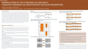 科学海报Manipulation of the Standard CFU-GM Assay Targeted Screening of Hematopoietic Myeloid Progenitors
 挂图Frequencies and Percentages of Mouse Immune Cell Types List of the frequencies of over 25 immune cell types in C57BL/6 mice
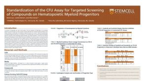


 沪公网安备31010102008431号