Rawat VPS et al. (SEP 2010)
Proceedings of the National Academy of Sciences of the United States of America 107 39 16946--51
The vent-like homeobox gene VENTX promotes human myeloid differentiation and is highly expressed in acute myeloid leukemia.
Recent data indicate that a variety of regulatory molecules active in embryonic development may also play a role in the regulation of early hematopoiesis. Here we report that the human Vent-like homeobox gene VENTX,a putative homolog of the Xenopus xvent2 gene,is a unique regulatory hematopoietic gene that is aberrantly expressed in CD34(+) leukemic stem-cell candidates in human acute myeloid leukemia (AML). Quantitative RT-PCR documented expression of the gene in lineage positive hematopoietic subpopulations,with the highest expression in CD33(+) myeloid cells. Notably,expression levels of VENTX were negligible in normal CD34(+)/CD38(-) or CD34(+) human progenitor cells. In contrast to this,leukemic CD34(+)/CD38(-) cells from AML patients with translocation t(8,21) and normal karyotype displayed aberrantly high expression of VENTX. Gene expression and pathway analysis demonstrated that in normal CD34(+) cells enforced expression of VENTX initiates genes associated with myeloid development and down-regulates genes involved in early lymphoid development. Functional analyses confirmed that aberrant expression of VENTX in normal CD34(+) human progenitor cells perturbs normal hematopoietic development,promoting generation of myeloid cells and impairing generation of lymphoid cells in vitro and in vivo. Stable knockdown of VENTX expression inhibited the proliferation of human AML cell lines. Taken together,these data extend our insights into the function of embryonic mesodermal factors in human postnatal hematopoiesis and indicate a role for VENTX in normal and malignant myelopoiesis.
View Publication
产品号#:
04434
04444
产品名:
MethoCult™ H4434 Classic
MethoCult™ H4434 Classic
Fiedler K et al. (JAN 2011)
Blood 117 4 1329--39
Neutrophil development and function critically depend on Bruton tyrosine kinase in a mouse model of X-linked agammaglobulinemia.
Bruton tyrosine kinase (Btk) is essential for B cell development and function and also appears to be important for myeloid cells. The bone marrow of Btk-deficient mice shows enhanced granulopoiesis compared with that of wild-type mice. In purified granulocyte-monocyte-progenitors (GMP) from Btk-deficient mice,the development of granulocytes is favored at the expense of monocytes. However,Btk-deficient neutrophils are impaired in maturation and function. Using bone marrow chimeras,we show that this defect is cell-intrinsic to neutrophils. In GMP and neutrophils,Btk plays a role in GM-CSF- and Toll-like receptor-induced differentiation. Molecular analyses revealed that expression of the lineage-determining transcription factors C/EBPα,C/EBPβ,and PU.1,depends on Btk. In addition,expression of several granule proteins,including myeloperoxidase,neutrophilic granule protein,gelatinase and neutrophil elastase,is Btk-dependent. In the Arthus reaction,an acute inflammatory response,neutrophil migration into tissues,edema formation,and hemorrhage are significantly reduced in Btk-deficient animals. Together,our findings implicate Btk as an important regulator of neutrophilic granulocyte maturation and function in vivo.
View Publication
产品号#:
03231
产品名:
MethoCult™ M3231
Bruserud &O et al. (MAR 2007)
Haematologica 92 3 332--41
Subclassification of patients with acute myelogenous leukemia based on chemokine responsiveness and constitutive chemokine release by their leukemic cells.
BACKGROUND AND OBJECTIVES: Chemokines are soluble mediators involved in angiogenesis,cellular growth control and immunomodulation. In the present study we investigated the effects of various chemokines on proliferation of acute myelogenous leukemia (AML) cells and constitutive chemokine release by primary AML cells. DESIGN AND METHODS: Native human AML cells derived from 68 consecutive patients were cultured in vitro. We investigated AML cell proliferation (3H-thymidine incorporation,colony formation),chemokine receptor expression,constitutive chemokine release and chemotaxis of normal peripheral blood mononuclear cells. RESULTS: Exogenous chemokines usually did not have any effect on AML blast proliferation in the absence of hematopoietic growth factors,but when investigating growth factor-dependent (interleukin 3 + granulocyte-macrophage colony-stimulating factor + stem cell factor) proliferation in suspension cultures the following patient subsets were identified: (i) patients whose cells showed chemokine-induced growth enhancement (8 patients); (ii) divergent effects on proliferation (15 patients); and (iii) no effect (most patients). These patient subsets did not differ in chemokine receptor expression,but,compared to CD34- AML cells,CD34+ cells showed higher expression of several receptors. Chemokines also increased the proliferation of clonogenic AML cells from the first subset of patients. Furthermore,a broad constitutive chemokine release profile was detected for most patients,and the following chemokine clusters could be identified: CCL2-4/CXCL1/8,CCL5/CXCL9-11 (possibly also CCL23) and CCL13/17/22/24/CXCL5 (possibly also CXCL6). Only the CCL2-4/CXCL1/8 cluster showed significant correlations between corresponding mRNA levels and NFkB levels/activation. The chemotaxis of normal immunocompetent cells for patients without constitutive chemokine release was observed to be decreased. INTERPRETATION AND CONCLUSIONS: Differences in chemokine responsiveness as well as chemokine release contribute to patient heterogeneity in AML. Patients with AML can be classified into distinct subsets according to their chemokine responsiveness and chemokine release profile.
View Publication
产品号#:
04434
04444
09600
09650
产品名:
MethoCult™ H4434 Classic
MethoCult™ H4434 Classic
StemSpan™ SFEM
StemSpan™ SFEM
Feng R et al. (MAR 2007)
Blood 109 5 2130--8
SDX-308, a nonsteroidal anti-inflammatory agent, inhibits NF-kappaB activity, resulting in strong inhibition of osteoclast formation/activity and multiple myeloma cell growth.
Multiple myeloma is characterized by increased osteoclast activity that results in bone destruction and lytic lesions. With the prolonged overall patient survival achieved by new treatment modalities,additional drugs are required to inhibit bone destruction. We focused on a novel and more potent structural analog of the nonsteroidal anti-inflammatory drug etodolac,known as SDX-308,and its effects on osteoclastogenesis and multiple myeloma cells. SDX-101 is another structural analog of etodolac that is already used in clinical trials for the treatment of B-cell chronic lymphocytic leukemia (B-CLL). Compared with SDX-101,a 10-fold lower concentration of SDX-308 induced potent (60%-80%) inhibition of osteoclast formation,and a 10- to 100-fold lower concentration inhibited multiple myeloma cell proliferation. Bone resorption was completely inhibited by SDX-308,as determined in dentin-based bone resorption assays. SDX-308 decreased constitutive and RANKL-stimulated NF-kappaB activation and osteoclast formation in an osteoclast cellular model,RAW 264.7. SDX-308 effectively suppressed TNF-alpha-induced IKK-gamma and IkappaB-alpha phosphorylation and degradation and subsequent NF-kappaB activation in human multiple myeloma cells. These results indicate that SDX-308 effectively inhibits multiple myeloma cell proliferation and osteoclast activity,potentially by controlling NF-kappaB activation signaling. We propose that SDX-308 is a promising therapeutic candidate to inhibit multiple myeloma growth and osteoclast activity and that it should receive attention for further study.
View Publication
产品号#:
04434
04444
产品名:
MethoCult™ H4434 Classic
MethoCult™ H4434 Classic
Welch JS et al. (FEB 2011)
Blood 117 8 2460--8
Rara haploinsufficiency modestly influences the phenotype of acute promyelocytic leukemia in mice.
RARA (retinoic acid receptor alpha) haploinsufficiency is an invariable consequence of t(15;17)(q22;q21) translocations in acute promyelocytic leukemia (APL). Retinoids and RARA activity have been implicated in hematopoietic self-renewal and neutrophil maturation. We and others therefore predicted that RARA haploinsufficiency would contribute to APL pathogenesis. To test this hypothesis,we crossed Rara(+/-) mice with mice expressing PML (promyelocytic leukemia)-RARA from the cathepsin G locus (mCG-PR). We found that Rara haploinsufficiency cooperated with PML-RARA,but only modestly influenced the preleukemic and leukemic phenotype. Bone marrow from mCG-PR(+/-) × Rara(+/-) mice had decreased numbers of mature myeloid cells,increased ex vivo myeloid cell proliferation,and increased competitive advantage after transplantation. Rara haploinsufficiency did not alter mCG-PR-dependent leukemic latency or penetrance,but did influence the distribution of leukemic cells; leukemia in mCG-PR(+/-) × Rara(+/-) mice presented more commonly with low to normal white blood cell counts and with myeloid infiltration of lymph nodes. APL cells from these mice were responsive to all-trans retinoic acid and had virtually no differences in expression profiling compared with tumors arising in mCG-PR(+/-) × Rara(+/+) mice. These data show that Rara haploinsufficiency (like Pml haploinsufficiency and RARA-PML) can cooperate with PML-RARA to influence the pathogenesis of APL in mice,but that PML-RARA is the t(15;17) disease-initiating mutation.
View Publication
产品号#:
03534
产品名:
MethoCult™ GF M3534
Arai S et al. (JUN 2011)
Blood 117 23 6304--14
Evi-1 is a transcriptional target of mixed-lineage leukemia oncoproteins in hematopoietic stem cells.
Ecotropic viral integration site-1 (Evi-1) is a nuclear transcription factor that plays an essential role in the regulation of hematopoietic stem cells. Aberrant expression of Evi-1 has been reported in up to 10% of patients with acute myeloid leukemia and is a diagnostic marker that predicts a poor outcome. Although chromosomal rearrangement involving the Evi-1 gene is one of the major causes of Evi-1 activation,overexpression of Evi-1 is detected in a subgroup of acute myeloid leukemia patients without any chromosomal abnormalities,which indicates the presence of other mechanisms for Evi-1 activation. In this study,we found that Evi-1 is frequently up-regulated in bone marrow cells transformed by the mixed-lineage leukemia (MLL) chimeric genes MLL-ENL or MLL-AF9. Analysis of the Evi-1 gene promoter region revealed that MLL-ENL activates transcription of Evi-1. MLL-ENL-mediated up-regulation of Evi-1 occurs exclusively in the undifferentiated hematopoietic population,in which Evi-1 particularly contributes to the propagation of MLL-ENL-immortalized cells. Furthermore,gene-expression analysis of human acute myeloid leukemia cases demonstrated the stem cell-like gene-expression signature of MLL-rearranged leukemia with high levels of Evi-1. Our findings indicate that Evi-1 is one of the targets of MLL oncoproteins and is selectively activated in hematopoietic stem cell-derived MLL leukemic cells.
View Publication
产品号#:
03434
03444
产品名:
MethoCult™ GF M3434
MethoCult™ GF M3434
Baba Y et al. (AUG 2006)
Journal of immunology (Baltimore,Md. : 1950) 177 4 2294--303
Constitutively active beta-catenin promotes expansion of multipotent hematopoietic progenitors in culture.
This study was designed to investigate one component of the Wnt/beta-catenin signaling pathway that has been implicated in stem cell self-renewal. Retroviral-mediated introduction of stable beta-catenin to primitive murine bone marrow cells allowed the expansion of multipotential c-Kit(low)Sca-1(low/-)CD19(-) CD11b/Mac-1(-)Flk-2(-)CD43(+)AA4.1(+)NK1.1(-)CD3(-)CD11c(-)Gr-1(-)CD45R/B220(+) cells in the presence of stromal cells and cytokines. They generated myeloid,T,and B lineage lymphoid cells in culture,but had no T lymphopoietic potential when transplanted. Stem cell factor and IL-6 were found to be minimal requirements for long-term,stromal-free propagation,and a beta-catenin-transduced cell line was maintained for 5 mo with these defined conditions. Although multipotential and responsive to many normal stimuli in culture,it was unable to engraft several types of irradiated recipients. These findings support previous studies that have implicated the canonical Wnt pathway signaling in regulation of multipotent progenitors. In addition,we demonstrate how it may be experimentally manipulated to generate valuable cell lines.
View Publication
产品号#:
03434
03444
产品名:
MethoCult™ GF M3434
MethoCult™ GF M3434
Nanua S et al. (MAR 2011)
Blood 117 13 3539--47
Activation of the unfolded protein response is associated with impaired granulopoiesis in transgenic mice expressing mutant Elane.
Severe congenital neutropenia (SCN) is an inborn disorder of granulopoiesis that in many cases is caused by mutations of the ELANE gene,which encodes neutrophil elastase (NE). Recent data suggest a model in which ELANE mutations result in NE protein misfolding,induction of endoplasmic reticulum (ER) stress,activation of the unfolded protein response (UPR),and ultimately a block in granulocytic differentiation. To test this model,we generated transgenic mice carrying a targeted mutation of Elane (G193X) reproducing a mutation found in SCN. The G193X Elane allele produces a truncated NE protein that is rapidly degraded. Granulocytic precursors from G193X Elane mice,though without significant basal UPR activation,are sensitive to chemical induction of ER stress. Basal and stress granulopoiesis after myeloablative therapy are normal in these mice. Moreover,inaction of protein kinase RNA-like ER kinase (Perk),one of the major sensors of ER stress,either alone or in combination with G193X Elane,had no effect on basal granulopoiesis. However,inhibition of the ER-associated degradation (ERAD) pathway using a proteosome inhibitor resulted in marked neutropenia in G193X Elane. The selective sensitivity of G913X Elane granulocytic cells to ER stress provides new and strong support for the UPR model of disease patho-genesis in SCN.
View Publication
Senn JJ et al. (SEP 2005)
The Journal of pharmacology and experimental therapeutics 314 3 972--9
Non-CpG-containing antisense 2'-methoxyethyl oligonucleotides activate a proinflammatory response independent of Toll-like receptor 9 or myeloid differentiation factor 88.
Oligonucleotides with a CpG" motif trigger a proinflammatory response through activation of Toll-like receptor 9 (TLR9) and are being studied to exploit these properties for use as adjuvants and cancer therapies. However�
View Publication
产品号#:
09600
09650
产品名:
StemSpan™ SFEM
StemSpan™ SFEM
Ragu C et al. (NOV 2010)
Blood 116 22 4464--73
The transcription factor Srf regulates hematopoietic stem cell adhesion.
Adhesion properties of hematopoietic stem cells (HSCs) in the bone marrow (BM) niches control their migration and affect their cell-cycle dynamics. The serum response factor (Srf) regulates growth factor-inducible genes and genes controlling cytoskeleton structures involved in cell spreading,adhesion,and migration. We identified a role for Srf in HSC adhesion and steady-state hematopoiesis. Conditional deletion of Srf in BM cells resulted in a 3-fold expansion of the long- and short-term HSCs and multipotent progenitors (MPPs),which occurs without long-term modification of cell-cycle dynamics. Early differentiation steps to myeloid and lymphoid lineages were normal,but Srf loss results in alterations in mature-cell production and severe thrombocytopenia. Srf-null BM cells also displayed compromised engraftment properties in transplantation assays. Gene expression analysis identified Srf target genes expressed in HSCs,including a network of genes associated with cell migration and adhesion. Srf-null stem cells and MPPs displayed impair expression of the integrin network and decreased adherence in vitro. In addition,Srf-null mice showed increase numbers of circulating stem and progenitor cells,which likely reflect their reduced retention in the BM. Altogether,our results demonstrate that Srf is an essential regulator of stem cells and MPP adhesion,and suggest that Srf acts mainly through cell-matrix interactions and integrin signaling.
View Publication

 EasySep™小鼠TIL(CD45)正选试剂盒


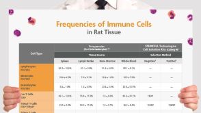 挂图Frequencies of Immune Cells in Rat Tissue Lists the estimated frequencies of more than 15 immune cell types in Sprague Dawley rats

 沪公网安备31010102008431号