Hartfield EM et al. (FEB 2014)
PLoS ONE 9 2 e87388
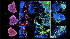
Physiological characterisation of human iPS-derived dopaminergic neurons
Human induced pluripotent stem cells (hiPSCs) offer the potential to study otherwise inaccessible cell types. Critical to this is the directed differentiation of hiPSCs into functional cell lineages. This is of particular relevance to research into neurological disease,such as Parkinson's disease (PD),in which midbrain dopaminergic neurons degenerate during disease progression but are unobtainable until post-mortem. Here we report a detailed study into the physiological maturation over time of human dopaminergic neurons in vitro. We first generated and differentiated hiPSC lines into midbrain dopaminergic neurons and performed a comprehensive characterisation to confirm dopaminergic functionality by demonstrating dopamine synthesis,release,and re-uptake. The neuronal cultures include cells positive for both tyrosine hydroxylase (TH) and G protein-activated inward rectifier potassium channel 2 (Kir3.2,henceforth referred to as GIRK2),representative of the A9 population of substantia nigra pars compacta (SNc) neurons vulnerable in PD. We observed for the first time the maturation of the slow autonomous pace-making (textless10 Hz) and spontaneous synaptic activity typical of mature SNc dopaminergic neurons using a combination of calcium imaging and electrophysiology. hiPSC-derived neurons exhibited inositol tri-phosphate (IP3) receptor-dependent release of intracellular calcium from the endoplasmic reticulum in neuronal processes as calcium waves propagating from apical and distal dendrites,and in the soma. Finally,neurons were susceptible to the dopamine neuron-specific toxin 1-methyl-4-phenylpyridinium (MPP+) which reduced mitochondrial membrane potential and altered mitochondrial morphology. Mature hiPSC-derived dopaminergic neurons provide a neurophysiologically-defined model of previously inaccessible vulnerable SNc dopaminergic neurons to bridge the gap between clinical PD and animal models.
View Publication
产品号#:
05850
05857
05870
05875
85850
85857
85870
85875
产品名:
mTeSR™1
mTeSR™1
Grunseich C et al. (OCT 2014)
Neurobiology of Disease 70 12--20
Stem cell-derived motor neurons from spinal and bulbar muscular atrophy patients.
Spinal and bulbar muscular atrophy (SBMA,Kennedy's disease) is a motor neuron disease caused by polyglutamine repeat expansion in the androgen receptor. Although degeneration occurs in the spinal cord and muscle,the exact mechanism is not clear. Induced pluripotent stem cells from spinal and bulbar muscular atrophy patients provide a useful model for understanding the disease mechanism and designing effective therapy. Stem cells were generated from six patients and compared to control lines from three healthy individuals. Motor neurons from four patients were differentiated from stem cells and characterized to understand disease-relevant phenotypes. Stem cells created from patient fibroblasts express less androgen receptor than control cells,but show androgen-dependent stabilization and nuclear translocation. The expanded repeat in several stem cell clones was unstable,with either expansion or contraction. Patient stem cell clones produced a similar number of motor neurons compared to controls,with or without androgen treatment. The stem cell-derived motor neurons had immunoreactivity for HB9,Isl1,ChAT,and SMI-32,and those with the largest repeat expansions were found to have increased acetylated ??-tubulin and reduced HDAC6. Reduced HDAC6 was also found in motor neuron cultures from two other patients with shorter repeats. Evaluation of stably transfected mouse cells and SBMA spinal cord showed similar changes in acetylated ??-tubulin and HDAC6. Perinuclear lysosomal enrichment,an HDAC6 dependent process,was disrupted in motor neurons from two patients with the longest repeats. SBMA stem cells present new insights into the disease,and the observations of reduced androgen receptor levels,repeat instability,and reduced HDAC6 provide avenues for further investigation of the disease mechanism and development of effective therapy. ?? 2014.
View Publication
产品号#:
05850
05857
05870
05875
85850
85857
85870
85875
产品名:
mTeSR™1
mTeSR™1
Bhinge A et al. (JUN 2014)
EMBO Journal 33 11 1271--1283
MiR-135b is a direct PAX6 target and specifies human neuroectoderm by inhibiting TGF-$\$/BMP signaling.
Several transcription factors (TFs) have been implicated in neuroectoderm (NE) development,and recently,the TF PAX6 was shown to be critical for human NE specification. However,microRNA networks regulating human NE development have been poorly documented. We hypothesized that microRNAs activated by PAX6 should promote NE development. Using a genomics approach,we identified PAX6 binding sites and active enhancers genome-wide in an in vitro model of human NE development that was based on neural differentiation of human embryonic stem cells (hESC). PAX6 binding to active enhancers was found in the proximity of several microRNAs,including hsa-miR-135b. MiR-135b was activated during NE development,and ectopic expression of miR-135b in hESC promoted differentiation toward NE. MiR-135b promotes neural conversion by targeting components of the TGF-β and BMP signaling pathways,thereby inhibiting differentiation into alternate developmental lineages. Our results demonstrate a novel TF-miRNA module that is activated during human neuroectoderm development and promotes the irreversible fate specification of human pluripotent cells toward the neural lineage.
View Publication

 EasySep™小鼠TIL(CD45)正选试剂盒


 27:19
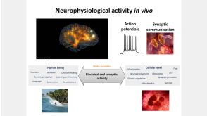
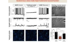
线上讲座BrainPhys™ Medium Supports the Physiological Activity of Neuronal Tissue in vitro发布日期: 07/22/2016
27:19
线上讲座BrainPhys™ Medium Supports the Physiological Activity of Neuronal Tissue in vitro发布日期: 07/22/2016

 沪公网安备31010102008431号