Ginis I et al. (JUN 2012)
Tissue engineering. Part C,Methods 18 6 453--63
Evaluation of bone marrow-derived mesenchymal stem cells after cryopreservation and hypothermic storage in clinically safe medium.
Achievements in tissue engineering using mesenchymal stem cells (MSC) demand a clinically acceptable off-the-shelf" cell therapy product. Efficacy of cryopreservation of human bone marrow-derived MSC in clinically safe animal product-free medium containing 2% 5% and 10% dimethyl sulfoxide (DMSO) was evaluated by measuring cell recovery viability apoptosis proliferation rate expression of a broad panel of MSC markers and osteogenic differentiation. Rate-controlled freezing in CryoStor media was performed in a programmable cell freezer. About 95% of frozen cells were recovered as live cells after freezing in CryoStor solutions with 5% and 10% DMSO followed by storage in liquid nitrogen for 1 month. Cell recovery after 5 months storage was 72% and 80% for 5% and 10% DMSO respectively. Measurements of caspase 3 activity demonstrated that 15.5% and 12.8% of cells after 1 month and 18.3% and 12.9% of cells after 5 months storage in 5% and 10% DMSO respectively were apoptotic. Proliferation of MSC recovered after cryopreservation was measured during 2 weeks post-plating. Proliferation rate was not compromised and was even enhanced. Cryopreservation did not alter expression of MSC markers. Quantitative analysis of alkaline phosphatase (ALP) activity ALP surface expression and Ca deposition in previously cryopreserved MSC and then differentiated for 3 weeks in osteogenic medium demonstrated the same degree of osteogenic differentiation as in unfrozen parallel cultures. Cell viability and functional parameters were analyzed in MSC after short-term storage at 4°C in HypoThermosol-FRS solution also free of animal products. Hypothermic storage for 2 and 4 days resulted in about 100% and 85% cell recovery respectively less than 10% of apoptotic cells and normal proliferation marker expression and osteogenic potential. Overall our results demonstrate that human MSC could be successfully cryopreserved for banking and clinical applications and delivered to the bedside in clinically safe protective reagents.
View Publication
Kitajima K et al. (JAN 2016)
Experimental hematology 44 1 10--68
GSK3$\$ activates the CDX/HOX pathway and promotes hemogenic endothelial progenitor differentiation from human pluripotent stem cells.
WNT/$\$-CATENIN signaling promotes the hematopoietic/endothelial differentiation of human embryonic stem cells and human induced pluripotent stem cells (hiPSCs). The transient addition of a GSK3$\$ (GSKi) has been found to facilitate in vitro endothelial cell differentiation from hESCs/hiPSCs. Because hematopoietic and endothelial cells are derived from common progenitors (hemogenic endothelial progenitors [HEPs]),we examined the effect of transient GSKi treatment on hematopoietic cell differentiation from hiPSCs. We found that transient GSKi treatment at the start of hiPSC differentiation induction altered the gene expression profile of the cells. Multiple CDX/HOX genes,which are expressed in the posterior mesoderm of developing embryos,were significantly upregulated by GSKi treatment. Further,inclusion of the GSKi in a serum- and stroma-free culture with chemically defined medium efficiently induced HEPs,and the HEPs gave rise to various lineages of hematopoietic and endothelial cells. Therefore,transient WNT/$\$-CATENIN signaling triggers activation of the CDX/HOX pathway,which in turn confers hemogenic posterior mesoderm identity to differentiating hiPSCs. These data enhance our understanding of human embryonic hematopoietic/endothelial cell development and provide a novel in vitro system for inducing the differentiation of hematopoietic cells from hiPSCs.
View Publication
产品类型:
产品号#:
04034
04044
05270
05275
85850
85857
产品名:
MethoCult™H4034 Optimum
MethoCult™H4034 Optimum
STEMdiff™ APEL™2 培养基
STEMdiff™ APEL™2 培养基
mTeSR™1
mTeSR™1
Ja KPMM et al. (FEB 2016)
Journal of cellular and molecular medicine 20 2 323--332
iPSC-derived human cardiac progenitor cells improve ventricular remodelling via angiogenesis and interstitial networking of infarcted myocardium.
We investigate the effects of myocardial transplantation of human induced pluripotent stem cell (iPSC)-derived progenitors and cardiomyocytes into acutely infarcted myocardium in severe combined immune deficiency mice. A total of 2 × 10(5) progenitors,cardiomyocytes or cell-free saline were injected into peri-infarcted anterior free wall. Sham-operated animals received no injection. Myocardial function was assessed at 2-week and 4-week post-infarction by using echocardiography and pressure-volume catheterization. Early myocardial remodelling was observed at 2-week with echocardiography derived stroke volume (SV) in saline (20.45 ± 7.36 $\$,P textless 0.05) and cardiomyocyte (19.52 ± 3.97 $\$,P textless 0.05) groups,but not in progenitor group (25.65 ± 3.61 $\$),significantly deteriorated as compared to sham control group (28.41 ± 4.41 $\$). Consistently,pressure-volume haemodynamic measurements showed worsening chamber dilation in saline (EDV: 23.24 ± 5.01 $\$,P textless 0.05; ESV: 17.08 ± 5.82 $\$,P textless 0.05) and cardiomyocyte (EDV: 26.45 ± 5.69 $\$,P textless 0.05; ESV: 18.03 ± 6.58 $\$,P textless 0.05) groups by 4-week post-infarction as compared to control (EDV: 15.26 ± 2.96 $\$; ESV: 8.41 ± 2.94 $\$). In contrast,cardiac progenitors (EDV: 20.09 ± 7.76 $\$; ESV: 13.98 ± 6.74 $\$) persistently protected chamber geometry against negative cardiac remodelling. Similarly,as compared to sham control (54.64 ± 11.37%),LV ejection fraction was preserved in progenitor group from 2-(38.68 ± 7.34%) to 4-week (39.56 ± 13.26%) while cardiomyocyte (36.52 ± 11.39%,P textless 0.05) and saline (35.34 ± 11.86%,P textless 0.05) groups deteriorated early at 2-week. Improvements of myocardial function in the progenitor group corresponded to increased vascularization (16.12 ± 1.49/mm(2) to 25.48 ± 2.08/mm(2) myocardial tissue,P textless 0.05) and coincided with augmented networking of cardiac telocytes in the interstitial space of infarcted zone.
View Publication
产品类型:
产品号#:
85850
85857
产品名:
mTeSR™1
mTeSR™1
Hawkins RD et al. (OCT 2011)
Cell Research 21 10 1393--1409
Dynamic chromatin states in human ES cells reveal potential regulatory sequences and genes involved in pluripotency.
Pluripotency,the ability of a cell to differentiate and give rise to all embryonic lineages,defines a small number of mammalian cell types such as embryonic stem (ES) cells. While it has been generally held that pluripotency is the product of a transcriptional regulatory network that activates and maintains the expression of key stem cell genes,accumulating evidence is pointing to a critical role for epigenetic processes in establishing and safeguarding the pluripotency of ES cells,as well as maintaining the identity of differentiated cell types. In order to better understand the role of epigenetic mechanisms in pluripotency,we have examined the dynamics of chromatin modifications genome-wide in human ES cells (hESCs) undergoing differentiation into a mesendodermal lineage. We found that chromatin modifications at promoters remain largely invariant during differentiation,except at a small number of promoters where a dynamic switch between acetylation and methylation at H3K27 marks the transition between activation and silencing of gene expression,suggesting a hierarchy in cell fate commitment over most differentially expressed genes. We also mapped over 50 000 potential enhancers,and observed much greater dynamics in chromatin modifications,especially H3K4me1 and H3K27ac,which correlate with expression of their potential target genes. Further analysis of these enhancers revealed potentially key transcriptional regulators of pluripotency and a chromatin signature indicative of a poised state that may confer developmental competence in hESCs. Our results provide new evidence supporting the role of chromatin modifications in defining enhancers and pluripotency.
View Publication
产品类型:
产品号#:
85850
85857
产品名:
mTeSR™1
mTeSR™1
Chan LY et al. (JAN 2013)
Biomaterials 34 2 382--392
Temporal application of topography to increase the rate of neural differentiation from human pluripotent stem cells.
Human pluripotent stem cells (hPSCs) are a promising cell source for tissue engineering and regenerative medicine,especially in the field of neurobiology. Neural differentiation protocols have been developed to differentiate hPSCs into specific neural cells,but these predominantly rely on biochemical cues. Recently,differentiation protocols have incorporated topographical cues to increase the total neuronal yield. However,the means by which these topographical cues improve neuronal yield remains unknown. In this study,we explored the effect of topography on the neural differentiation of hPSC by quantitatively studying the changes in marker expression at a transcript and protein level. We found that 2 ??m gratings increase the rate of neural differentiation,and that an additional culture period of 2 ??m gratings in the absence of neurotrophic signals can improve the neural differentiation of hPSCs. We envisage that this work can be incorporated into future differentiation protocols to decrease the differentiation period as well as the biochemical signals added,thus generating hPSC-derived neural cells in a more cost effective and efficient manner. ?? 2012 Elsevier Ltd.
View Publication
产品类型:
产品号#:
85850
85857
产品名:
mTeSR™1
mTeSR™1
Yang S-L et al. (DEC 2012)
Protein & cell 3 12 934--942
Compound screening platform using human induced pluripotent stem cells to identify small molecules that promote chondrogenesis.
Articular cartilage,which is mainly composed of collagen II,enables smooth skeletal movement. Degeneration of collagen II can be caused by various events,such as injury,but degeneration especially increases over the course of normal aging. Unfortunately,the body does not fully repair itself from this type of degeneration,resulting in impaired movement. Microfracture,an articular cartilage repair surgical technique,has been commonly used in the clinic to induce the repair of tissue at damage sites. Mesenchymal stem cells (MSC) have also been used as cell therapy to repair degenerated cartilage. However,the therapeutic outcomes of all these techniques vary in different patients depending on their age,health,lesion size and the extent of damage to the cartilage. The repairing tissues either form fibrocartilage or go into a hypertrophic stage,both of which do not reproduce the equivalent functionality of endogenous hyaline cartilage. One of the reasons for this is inefficient chondrogenesis by endogenous and exogenous MSC. Drugs that promote chondrogenesis could be used to induce self-repair of damaged cartilage as a non-invasive approach alone,or combined with other techniques to greatly assist the therapeutic outcomes. The recent development of human induced pluripotent stem cell (iPSCs),which are able to self-renew and differentiate into multiple cell types,provides a potentially valuable cell resource for drug screening in a more relevant" cell type. Here we report a screening platform using human iPSCs in a multi-well plate format to identify compounds that could promote chondrogenesis."
View Publication
产品类型:
产品号#:
85850
85857
产品名:
mTeSR™1
mTeSR™1
Zhu H et al. (OCT 2013)
Nucleic Acids Research 41 19 e180
Baculoviral transduction facilitates TALEN-mediated targeted transgene integration and Cre/LoxP cassette exchange in human-induced pluripotent stem cells
Safety and reliability of transgene integration in human genome continue to pose challenges for stem cell-based gene therapy. Here,we report a baculovirus-transcription activator-like effector nuclease system for AAVS1 locus-directed homologous recombination in human induced pluripotent stem cells (iPSCs). This viral system,when optimized in human U87 cells,provided a targeted integration efficiency of 95.21% in incorporating a Neo-eGFP cassette and was able to mediate integration of DNA insert up to 13.5 kb. In iPSCs,targeted integration with persistent transgene expression was achieved without compromising genomic stability. The modified iPSCs continued to express stem cell pluripotency markers and maintained the ability to differentiate into three germ lineages in derived embryoid bodies. Using a baculovirus-Cre/LoxP system in the iPSCs,the Neo-eGFP cassette at the AAVS1 locus could be replaced by a Hygro-mCherry cassette,demonstrating the feasibility of cassette exchange. Moreover,as assessed by measuring γ-H2AX expression levels,genome toxicity associated with chromosomal double-strand breaks was not detectable after transduction with moderate doses of baculoviral vectors expressing transcription activator-like effector nucleases. Given high targeted integration efficiency,flexibility in transgene exchange and low genome toxicity,our baculoviral transduction-based approach offers great potential and attractive option for precise genetic manipulation in human pluripotent stem cells.
View Publication
产品类型:
产品号#:
07923
85850
85857
产品名:
Dispase (1 U/mL)
mTeSR™1
mTeSR™1
Ko J-YY et al. (APR 2014)
Biomaterials 35 11 3571--3581
In vitro chondrogenesis and in vivo repair of osteochondral defect with human induced pluripotent stem cells.
The purpose of this study was to investigate the chondrogenic features of human induced pluripotent stem cells (hiPSCs) and examine the differences in the chondrogenesis between hiPSCs and human bone marrow-derived MSCs (hBMMSCs). Embryoid bodies (EBs) were formed from undifferentiated hiPSCs. After EBs were dissociated into single cells,chondrogenic culture was performed in pellets and alginate hydrogel. Chondro-induced hiPSCs were implanted in osteochondral defects created on the patellar groove of immunosuppressed rats and evaluated after 12 weeks. The ESC markers NANOG,SSEA4 and OCT3/4 disappeared while the mesodermal marker BMP-4 appeared in chondro-induced hiPSCs. After 21 days of culture,greater glycosaminoglycan contents and better chondrocytic features including lacuna and abundant matrix formation were observed from chondro-induced hiPSCs compared to chondro-induced hBMMSCs. The expression of chondrogenic markers including SOX-9,type II collagen,and aggrecan in chondro-induced hiPSCs was comparable to or greater than chondro-induced hBMMSCs. A remarkably low level of hypertrophic and osteogenic markers including type X collagen,type I collagen and Runx-2 was noted in chondro-induced hiPSCs compared to chondro-induced hBMMSCs. hiPSCs had significantly greater methylation of several CpG sites in COL10A1 promoter than hBMMSCs in either undifferentiated or chondro-induced state,suggesting an epigenetic cause of the difference in hypertrophy. The defects implanted with chondro-induced hiPSCs showed a significantly better quality of cartilage repair than the control defects,and the majority of cells in the regenerated cartilage consisted of implanted hiPSCs. ?? 2014 Elsevier Ltd.
View Publication
产品类型:
产品号#:
85850
85857
产品名:
mTeSR™1
mTeSR™1
Lian X et al. (NOV 2014)
Stem cell reports 3 5 804--816
Efficient differentiation of human pluripotent stem cells to endothelial progenitors via small-molecule activation of WNT signaling.
Human pluripotent stem cell (hPSC)-derived endothelial cells and their progenitors may provide the means for vascularization of tissue-engineered constructs and can serve as models to study vascular development and disease. Here,we report a method to efficiently produce endothelial cells from hPSCs via GSK3 inhibition and culture in defined media to direct hPSC differentiation to CD34(+)CD31(+) endothelial progenitors. Exogenous vascular endothelial growth factor (VEGF) treatment was dispensable,and endothelial progenitor differentiation was β-catenin dependent. Furthermore,by clonal analysis,we showed that CD34(+)CD31(+)CD117(+)TIE-2(+) endothelial progenitors were multipotent,capable of differentiating into calponin-expressing smooth muscle cells and CD31(+)CD144(+)vWF(+)I-CAM1(+) endothelial cells. These endothelial cells were capable of 20 population doublings,formed tube-like structures,imported acetylated low-density lipoprotein,and maintained a dynamic barrier function. This study provides a rapid and efficient method for production of hPSC-derived endothelial progenitors and endothelial cells and identifies WNT/β-catenin signaling as a primary regulator for generating vascular cells from hPSCs.
View Publication
Generation of GFAP::GFP astrocyte reporter lines from human adult fibroblast-derived iPS cells using zinc-finger nuclease technology.
Astrocytes are instrumental to major brain functions,including metabolic support,extracellular ion regulation,the shaping of excitatory signaling events and maintenance of synaptic glutamate homeostasis. Astrocyte dysfunction contributes to numerous developmental,psychiatric and neurodegenerative disorders. The generation of adult human fibroblast-derived induced pluripotent stem cells (iPSCs) has provided novel opportunities to study mechanisms of astrocyte dysfunction in human-derived cells. To overcome the difficulties of cell type heterogeneity during the differentiation process from iPSCs to astroglial cells (iPS astrocytes),we generated homogenous populations of iPS astrocytes using zinc-finger nuclease (ZFN) technology. Enhanced green fluorescent protein (eGFP) driven by the astrocyte-specific glial fibrillary acidic protein (GFAP) promoter was inserted into the safe harbor adeno-associated virus integration site 1 (AAVS1) locus in disease and control-derived iPSCs. Astrocyte populations were enriched using Fluorescence Activated Cell Sorting (FACS) and after enrichment more than 99% of iPS astrocytes expressed mature astrocyte markers including GFAP,S100$\$,NFIA and ALDH1L1. In addition,mature pure GFP-iPS astrocytes exhibited a well-described functional astrocytic activity in vitro characterized by neuron-dependent regulation of glutamate transporters to regulate extracellular glutamate concentrations. Engraftment of GFP-iPS astrocytes into rat spinal cord grey matter confirmed in vivo cell survival and continued astrocytic maturation. In conclusion,the generation of GFAP::GFP-iPS astrocytes provides a powerful in vitro and in vivo tool for studying astrocyte biology and astrocyte-driven disease pathogenesis and therapy.
View Publication

 EasySep™小鼠TIL(CD45)正选试剂盒


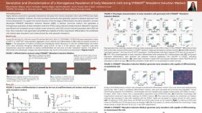 科学海报Generation and Characterization of a Homogenous Population of Early Mesoderm Cells Using STEMdiff Mesoderm Induction Medium

 沪公网安备31010102008431号